Articles
FluoroFinder News & Updates
From flow cytometry research and experimental design trends to FluoroFinder tool updates and industry applications, we explore it all in our blog.
Intravital Microscopy – Explained
Intravital microscopy has vast potential to reveal hidden cellular mechanisms Intravital microscopy (IVM) is a term used to describe the direct visualization of cells and tissues within a living organism. It was first reported in the 17th century, shortly after...
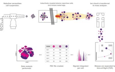
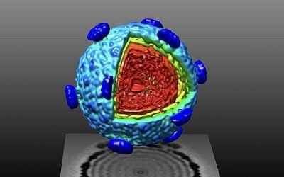
Mass Cytometry – Explained
The use of mass cytometry is growing and includes its adoption for clinical applications Mass cytometry is a variation of flow cytometry that uses antibodies labeled with metal tags rather than fluorophores. Also known as CyTOF® flow cytometry, in deference to...
Choosing the Right Fluorescent Immunoassay for Your Research
Fluorescent immunoassays meet different experimental needs When immunoassays such as western blot and enzyme-linked immunosorbent assay (ELISA) were first reported, they typically produced either a radiometric or chromogenic signal. However, many common...
Staining Strategies for Intracellular Flow Cytometry
By measuring intracellular proteins with flow cytometry, researchers can better understand cell signaling and functional responses in conditions of health and disease Flow cytometry is widely used for identifying different cell types based on the expression of...
Guide To Selecting Fluorophores for ICC and IHC
Choosing the right fluorophores for microscopy-based research is critical for accurate results Using fluorescent reagents for microscopy-based research offers several advantages. Not only does fluorescent detection enable multiplexing, which can be especially useful...
Flow Cytometry Troubleshooting Guide
Common problems can often be addressed by optimizing experimental design Flow cytometry is a technique for analyzing individual cells in suspension. It works by using a stream of fluid to direct the cells in single file past an interrogation point, where one or...
Viability Dye Selection Guide
Viability dyes are critical controls for proper flow analysis. The presence of dead cells can result in unwanted autofluorescence, cell aggregation, non-specific antibody binding, and a compromised capacity for RNA/DNA to proliferate in downstream applications. These...
Tips to Optimize Your Fluorescent Western Blot Protocol
Fluorescent western blot detection can offer many advantages provided protocols are carefully optimized While it was once common for researchers to use enzyme-labeled antibodies and chemiluminescent substrates to develop western blots to film, there has been a...
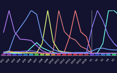
Panel Design for Spectral Flow Cytometry
Spectral cytometry offers increased flexibility for fluorophore selection but researchers should still apply best practices for panel design. It has been almost 20 years since spectral cytometry was first described by the Robinson group at Purdue University...
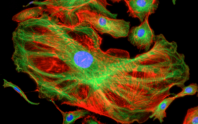
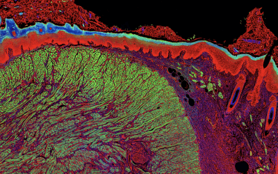
Picture Perfect: Capturing a High-Quality Fluorescent Microscopy Image
Protocol optimization is essential for clear, reproducible results Fluorescence microscopy uses fluorophore-labeled antibodies and other fluorescent reagents to visualize targets of interest in cells and tissues. Experiments can be as simple as detecting one...
Antigen Density for Flow Cytometry
Understanding the biological density of proteins or antigens expressed by each individual cell is an imperative component of all cell-based analytical methods. The comparative change in protein expression, also called antigen density, is indicative of the...
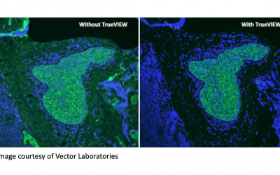
Tips to Minimize Autofluorescence
Reducing autofluorescence is critical in fluorescence-based research Techniques such as fluorescence microscopy, flow cytometry, and western blotting often rely on the use of fluorophore-labeled antibodies. The main reason for this is that fluorescence-based...
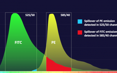
Compensation in Flow Cytometry
Fluorescent cellular analytical technologies allow us to “see” beyond what was historically possible with histological stains or morphological scatter profiles. In the early days, microscopy employed excitation sources like arc lamps, isolating specific wavelengths of...
Tips for Fluorophore Selection
Choosing the right fluorophores is critical for reliable results Fluorescent detection offers significant advantages, including multiplexing capability, superior sensitivity, and a broader dynamic linear range compared to other detection methods. However, the...
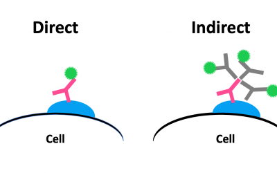
Direct vs Indirect Detection in Microscopy
Understanding the advantages and disadvantages of different immunodetection strategies is important to achieve accurate results. Microscopy-based techniques such as immunocytochemistry (ICC) and immunohistochemistry (IHC) often use fluorophore or enzyme-labeled...
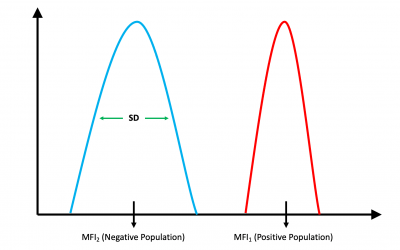
Stain Index for Flow Cytometry – Explained
When designing a flow cytometry experiment, it is important to account for the relative brightness of each fluorescent label on your specific instrument. Ideally, brighter fluorophores should be assigned to weakly expressed markers, while dimmer fluorophores should...
Confocal vs Super-Resolution Microscopy
Advanced microscopy techniques enable deeper imaging Advanced microscopy platforms are becoming more widespread for the depth of information they provide. Among these newer modalities, confocal microscopy has risen in popularity for imaging thick...

5 Recipes for Flow Cytometry Buffers
Whether preformulated or made in-house, buffers are integral to flow cytometry success Flow cytometry allows researchers to characterize individual cells using fluorophore-labeled antibodies for detecting targets of interest. Sample preparation, antibody...
Computational Microscopy
Non-conventional optics and advanced algorithms are extending the capabilities of classic microscopy. For centuries, researchers have tried to improve the resolution and sensitivity of optical microscopy. Over time, advances in fluorescence imaging modalities...
Take Control of Your Flow Cytometry Assay
Flow cytometry controls must address multiple sources of variation Flow cytometry requires more controls than other immunoassay techniques because it accounts for a greater number of potential sources of variation. In addition to experimental controls that...
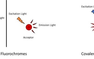
Tandem Dyes
The commercial availability of antibody-linked fluorescent dyes has expanded rapidly over the past decade. Where researchers were once limited to a handful of common dyes (FITC, PE, APC, PerCP, etc.), they can now choose from an extensive list of fluorescent dye lines...
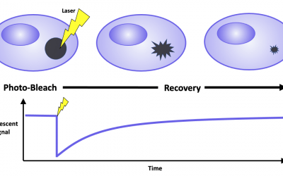
Fluorescence Recovery After Photobleaching
Fluorescence recovery after photobleaching is a technique for characterizing the mobility of cellular molecules Fluorescence recovery after photobleaching (FRAP), also known as fluorescence redistribution after photobleaching, is a microscopy-based technique...
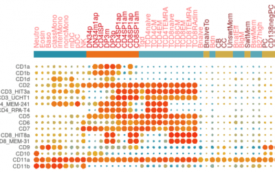
Optimizing Your Immunophenotyping Assay
Antibody conjugate panels must be designed with care to ensure accurate results Immunophenotyping is a flow cytometry-based technique which uses fluorophore-labeled antibodies for identifying different cell types within a heterogeneous population. While some cell...

Advances in Microscopy
Microscopy is comprised of many innovative technologies and remains at the forefront of scientific research. Since the invention of the microscope in the late 16th century, there has been a continual push to produce instruments with higher resolution, faster...

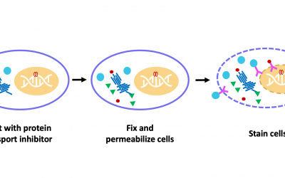
Guide to Fixation and Permeabilization
Fixation and permeabilization are key protocol steps for several core immunoassay techniques Immunoassay techniques such as flow cytometry, immunocytochemistry (ICC), and immunohistochemistry (IHC) often require that samples be fixed and/or permeabilized prior to...
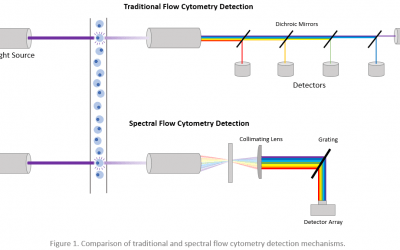
Roundup of Spectral Flow Cytometers
The use of spectral flow cytometry is increasing due to the many benefits it affords. Spectral flow cytometry is a relatively new technology that was developed to push the boundaries of traditional flow cytometry. First described in 2004 by researchers at Purdue...
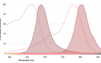
Near-Infrared Fluorophores For Fluorescence Imaging
Near-infrared fluorophores offer several advantages for imaging applications. A major advantage of fluorescent detection is that it enables multiple antibodies to be combined in the same experiment. Using techniques such as immunocytochemistry (ICC) and...

Fantastic Beasts and How To Image Them
Fluorescence microscopy is seeing increased utility for imaging organisms While fluorescence microscopy has long been employed for imaging plated mammalian cells, its use for visualizing organisms is flourishing. In recent years, winning images in Nikon’s annual Small...
Preparing a Single-Cell Suspension for Flow Cytometry
High viabilities, well-preserved antigens, and minimal clumping underpin flow cytometry success Unlike bulk population analysis techniques such as ELISA and Western blot, flow cytometry provides information about individual cells. Although microscopy does this as...

OMIPs
What are OMIPs? Optimized multicolor immunofluorescence panels, commonly known as OMIPs, are highly validated sets of antibodies and reagents that are published, peer-reviewed, and compiled to serve as a resource to the scientific community. OMIP publications often...
Advances in Western Blot Technologies
New approaches to Western blotting techniques overcome performance limitations For over 40 years, researchers have used Western blot for protein identification. Not only does Western Blot provide confirmation that a target of interest is present in a sample, this...
Expansion Microscopy
Expansion Microscopy physically expands biological specimens enabling higher resolution imaging First described in 2015, expansion microscopy is an imaging technique that improves on the resolution of conventional light microscopy by physically expanding the...
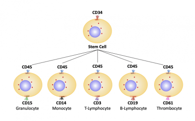
Flow Cytometry in Stem Cell Research and Therapy
Identifying distinct stem cell types and monitoring their differentiation is essential for developing novel therapies Stem cell research originated in the 1950s when it was discovered that mice which had received a lethal dose of radiation could recover...
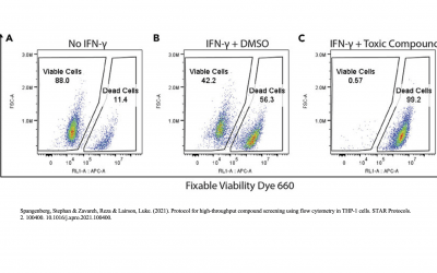
Excluding Dead Cells With a Cell Viability Fluorescent Dye
Excluding dead cells from flow cytometry analysis with a cell viability fluorescent dye is critical for producing accurate results The presence of dead cells in samples that will be analyzed by flow cytometry can affect antibody staining and lead to inaccurate...
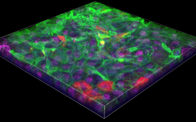
Newsletter: Imaging and Analysis of 3D Cell Cultures
Compared to traditional monolayers, 3D cell cultures more closely resemble physiological conditions 2-dimensional (2D) cell cultures consisting of monolayers attached to a flat surface have been a mainstay of scientific research for decades. However, cells grown in...

Newsletter: Considerations for Spectral Flow Cytometry Panel Design
Spectral flow cytometry offers several advantages over conventional flow cytometry but may be more affected by spreading error Spectral flow cytometry was first described in 2004 by the Robinson group at Purdue University Cytometry Laboratories. Since then, several...
Newsletter: Confocal Microscopy and Related Technologies
Confocal microscopy offers several advantages over traditional widefield microscopy and has evolved to meet many different research needs Originally developed by Marvin Minsky in the 1950s to address the problem of light scatter, confocal microscopy has become an...
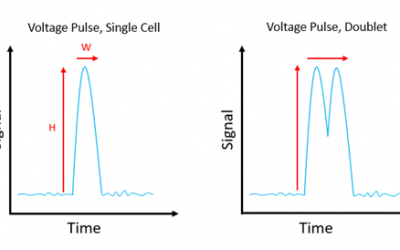
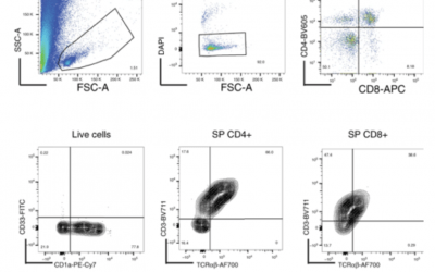
Newsletter: Distinguishing Cell-Cell Complexes from Singlet Cells
Failure to correctly identify cell-cell complexes can lead to data being misinterpreted and compromise the purity of sorted cell populations Multiparametric flow cytometry allows researchers to detect different cell types within a heterogeneous population. When...

Newsletter: Antibody Selection Guidelines
Choosing the right antibody for the intended application and sample type is critical to producing reliable results Antibodies are essential tools for applications ranging from flow cytometry, immunocytochemistry (ICC), and immunohistochemistry (IHC) to Western...
Newsletter: Comparison of Flow Cytometry Analysis Software
Software packages for viewing and analyzing FCS data files offer different features and benefits Data analysis represents a major source of variation in flow cytometry. Not only is the analysis process highly prone to user subjectivity, but disparities may arise due...