Fluorescent western blot detection can offer many advantages provided protocols are carefully optimized
While it was once common for researchers to use enzyme-labeled antibodies and chemiluminescent substrates to develop western blots to film, there has been a shift in recent years. The introduction of modern digital imaging instruments, combined with advances in antibody labeling, have led many labs to transition to fluorescent western blot detection. Here, Abcam and Azure Biosystems comment on the advantages of fluorescent western blotting and share tips for protocol optimization.
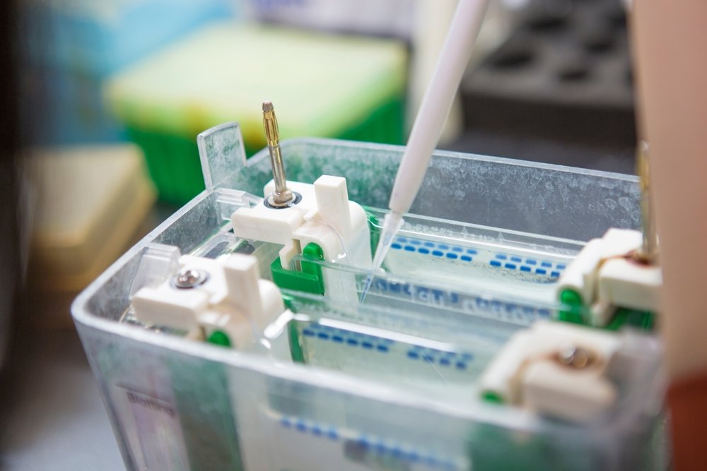
The Fluorescent Western Blot Protocol
A standard fluorescent western blot protocol mirrors the steps of conventional western blotting. First, the samples are separated by polyacrylamide gel electrophoresis (PAGE) and transferred to a membrane. Next, the membrane is blocked to prevent non-specific antibody binding and the target of interest is detected. Detection can either be direct, using labeled primary antibodies, or indirect, using unlabeled primary antibodies and labeled secondary antibodies. However, while chemiluminescent detection involves capturing an image of blots using film or a digital imaging system, fluorescent detection requires the use of an imaging instrument capable of measuring fluorescent signals.
Advantages of Fluorescent Western Blot Detection
“A main advantage of fluorescent western blot detection is that it allows for multiplexing,” reports Jade Fee, Application Scientist at Azure Biosystems. “This can be especially useful when sample material is limited or there is a need to detect proteins that have a similar molecular weight.” Fee explains that with chemiluminescent methods, detecting similar-sized bands requires the blot to be stripped and re-probed. In contrast, fluorescent detection allows researchers to simultaneously measure up to four different targets using modern imaging systems such as Sapphire FL™ Biomolecular Imager from Azure Biosystems.
“Multiplexing in fluorescent western blotting allows you to quantify relative protein abundance,” adds Silvia Sbacchi, Team Lead, Immunohistochemistry and Western Blot at Abcam. “For instance, you can compare the abundance of a phosphorylated form of your protein of interest to the total amount of protein. You can also perform normalization of band intensity with an internal control in the same blot, without the inconvenience of stripping and re-probing.”
In comparison to chemiluminescent methods, fluorescent western blot detection can improve experimental reproducibility due to the increased stability of fluorescent signals. Additionally, the linear relationship between fluorescence and the quantity of antibody bound to the blot allows for more precise quantification of protein targets. However, the limited detection sensitivity of fluorescent western blotting can pose challenges when detecting antigens with low abundance.
Tips for Success
As with any immunoassay, fluorescent western blotting requires careful preparation and optimization to generate accurate, reproducible results. Here are some tips to maximize the efficacy of your fluorescent western blot experiments:
-
Select Compatible Fluorophores for Your Imaging System
Understanding the lasers and detectors of your imaging system is critical for fluorophore selection. FluoroFinder’s Spectra Viewer enables you to visualize the excitation and emission spectra of over 1,100 fluorophores in the context of your specific laser and filter configuration. “If an antibody is not available labeled with your chosen fluorophore, it is now relatively straightforward to perform conjugations in-house,” comments Sbacchi. “Our Lightning-Link® kits let you label antibodies in as little as 15-minutes and come with over 20 different fluorophore options.”
-
Avoid Using Bromophenol Blue for Loading
While bromophenol blue is often included as a loading dye in sample buffers, it will autofluoresce once transferred to the membrane. “When using bromophenol blue, we recommend cutting the dye front from the gel prior to transfer,” says Sbacchi. “However, if this risks removing a low molecular weight protein target, researchers should consider switching to a different sample buffer.”
-
Choose a Suitable Molecular Weight Marker
Pre-stained molecular weight markers allow researchers to validate successful protein separation and transfer, as well as estimate the size of detected bands. However, many visible dyes included in common protein ladders have fluorescent properties, which can interfere with the detection of target proteins. To address this, it is recommended to use a protein ladder specifically designed for fluorescent western blotting. The iBright™ Prestained Protein Ladders and PageRuler™ Prestained NIR Protein Ladders both contain fluorophore-labeled proteins and colored markers visible to the naked eye.
-
Take Caution with Membrane Selection
Polyvinyl difluoride (PVDF) are less ideal for fluorescent western blotting because of their tendency to autofluoresce. “We suggest using either nitrocellulose or a low-fluorescence PVDF membrane for fluorescent detection,” says Sbacchi. “We also recommend handling the membrane with care to avoid scratching or creasing and using a pencil, not a pen, if you need to mark the membrane as many inks can autofluoresce.”
-
Consider Various Blocking Agents
While milk is widely used as a blocking agent, it often contains undissolved particles that can settle on the membrane and create fluorescent artifacts. Additionally, milk is often diluted in a buffer with a low concentration of detergent, a source of potential autofluorescence. However, there are commercial blocking agents that can be used in substitution of milk. The Azure Fluorescent Blot Blocking Buffer was specifically designed for fluorescent detection. “This blocking buffer is optimized for use with the Azure Imaging Systems and formulated to reduce background noise and stabilize fluorescent signals,” comments Fee.
-
Select the Right Antibodies
It is important to select antibodies validated for western blotting and titrate your selected products to maximize the signal-to-noise ratio. With over three million antibodies, our Product Search tool is a useful resource for discovering the latest fluorophore-labeled antibodies available for your antigens of interest. “Fluorescent western blots often require different antibody concentrations than chemiluminescent western blots,” cautions Sbacchi. “Researchers should therefore never assume that the same experimental conditions can be applied across both detection methods.” When it comes to storage, fluorophore-labeled antibodies should always be kept in the dark to avoid photobleaching.
-
Considerations for Multiplexing
Multiplexing can introduce unwanted cross-reactivities, particularly when using indirect detection. We recommend selecting primary antibodies from distinct and distantly-related species or of different isotypes and secondary antibodies that are highly cross-adsorbed. It is also recommended to select spectrally-distinct fluorophores to minimize unwanted background. FluoroFinder’ Spectra Viewer allows you to visualize fluorophore-labeled antibodies available for your targets of interest to help minimize spectral overlap, and subsequently unwanted background.
Supporting Your Fluorescent Western Blot
FluoroFinder has developed a suite of tools for reagent selection and experiment design. Our Spectra Viewer can help you identify fluorophores compatible with your imaging system, and our Product Search tool can help you find antibodies for your specific target of interest that are validated for western blotting.
Our partners also offer a wealth of resources. Explore Azure’s western blot guide to learn how to transition from chemiluminescent to fluorescent western blot detection. We also recommend Azure’s application notes on performing 3-color western blots with the Azure 600 Imager and 4-color western blots with the Sapphire FL™ Biomolecular Imager, as well as Abcam’s fluorescent western blotting resource page and guide for using their IRDye® secondary antibodies.
Sign-up for our eNewsletter to receive regular updates and resources about fluorescent western blotting, flow cytometry, and fluorescent microscopy.


