Fluorescent immunoassays meet different experimental needs
When immunoassays such as western blot and enzyme-linked immunosorbent assay (ELISA) were first reported, they typically produced either a radiometric or chromogenic signal. However, many common immunoassay techniques now use fluorescent detection for the advantages that it provides. These include higher sensitivity, a broader dynamic linear range, and more stable signals than traditional readouts, as well as greater quantitation accuracy and the capacity for multiplexing. But how do you decide which fluorescent immunoassay is right for your research? We spoke with Bio-Techne, Biotium, and Vector Laboratories, who shared some insights to help guide your decision.
Western Blot
Western blot is a technique for detecting proteins in solution (usually a cell lysate or tissue homogenate) following their separation by polyacrylamide gel electrophoresis (PAGE) and subsequent transfer to a nitrocellulose or PVDF membrane. A main advantage of western blot is that it requires only small amounts of antibody reagents to detect the target of interest. On the flipside, western blot has limited throughput, although this has recently been improved with automated platforms such as Bio-Techne’s Simple Western™ Automated Western Blot Systems. Common applications of western blot include evaluating protein abundance, monitoring kinase activity or post-translational modifications, and studying protein-protein interactions. In addition, western blot is widely used for antibody validation purposes.
“Using fluorophore conjugated antibodies for western blot offers a greater linear dynamic range than chemiluminescence, enabling a higher sensitivity threshold that can increase researchers’ chances of detecting low abundance proteins,” notes Emily Cartwright, Ph.D., Senior Product Marketing Specialist at Bio-Techne. “It is also possible with fluorescent detection to simultaneously probe for multiple targets, thus saving time spent on stripping and re-probing the membrane. Another important advantage of fluorescent western blot detection is that the signals are stable for weeks to months, compared to just 6-24 hours with chemiluminescence, which is ideal for digital imaging. Through our Novus Biologicals brand, Bio-Techne offers more than 70,000 western blot-validated antibodies backed by our 100% guarantee.”
ELISA
ELISA is a plate-based method for detecting soluble analytes. It can be configured in several different ways, of which the sandwich ELISA format is most often used due to its enhanced specificity. Advantages of ELISA include its high sensitivity, ease of use, and capacity to provide quantitative results. It is also readily automated to support high throughput applications. The only real limitation of ELISA is the fact that it is a bulk population analysis method, meaning it cannot provide information at the single-cell level. ELISA has an almost infinite number of uses, spanning small molecule screening during drug discovery and development to monitoring of disease biomarkers for diagnostic and clinical applications.
Currently, many ELISAs rely on antibodies labeled with enzymes such as horseradish peroxidase (HRP) or alkaline phosphatase (AP) for detection. These serve to convert a matched substrate to a colored or luminescent reaction product. However, ELISAs can also use antibodies labeled with fluorophores, when they are sometimes referred to as fluorescence-linked immunosorbent assays (FLISAs). When developing a FLISA, it is important to match the fluorophore labels to the reader’s lasers and detectors, and to use opaque black microplates to minimize well-to-well cross-talk and unwanted background fluorescence.
Immunohistochemistry (IHC)
IHC is the process of immunostaining tissue sections, which may either be frozen or formalin-fixed and paraffin-embedded (FFPE). Critically, unlike techniques such as western blot and ELISA, IHC provides information about protein localization, which is useful for understanding how antigen distribution differs between conditions of health and disease. The first reported IHC study used fluorescein isothiocyanate (FITC) labeled antibodies to identify pneumococcal antigens in infected tissues. Now, IHC is used for applications that include identifying novel biomarkers, diagnosing conditions such as cancer, inflammatory disease, and viral infections, and monitoring a patient’s response to drug treatment.
There are numerous factors to consider when performing fluorescent IHC detection, especially when working with FFPE tissue. “Cross-links introduced by formalin fixation can be a major source of autofluorescence,” explains Erika Leonard, Director of R&D at Vector Laboratories. “Additionally, autofluorescence can come from sample components such as collagen, elastin, and NADH. To address this problem, we developed the Vector® TrueVIEW™ Autofluorescence Quencher Kit, which diminishes autofluorescence from non-lipofuscin sources to dramatically improve the signal-to-noise ratio.” Another critical consideration is the type of mounting media that is used. “It is recommended that researchers select a mounting media that is optimized for immunofluorescence applications,” cautions Leonard. “Our range of VECTASHIELD® Antifade Mounting Media is designed to protect fluorophores from photobleaching, ensuring fluorescent signals are retained for both image acquisition and specimen archiving.”
Immunocytochemistry (ICC)
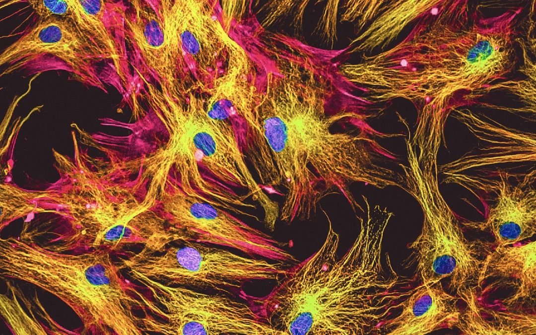
ICC uses antibodies for visualizing analytes of interest on and/or within cells. The types of samples that are studied include adherent cell lines, primary cells, and stem cell cultures, as well as suspension cells such as blood and swab samples. ICC is also applied for long-term live cell imaging studies. An advantage of ICC is that, like IHC, it provides spatial information about the targets being investigated. But, also like IHC, ICC faces challenges associated with autofluorescence, which can be compounded by the choice of culture vessel.
Coverglass cultureware is widely used for ICC and is recommended over chambered coverglasses if the cells will be fixed and permeabilized. This is because fixation and permeabilization can cause the chambers to leak, leading to experimental artifacts from antibodies entering neighboring wells. A low cost alternative to coverglass cultureware is to culture the cells on glass coverslips coated with poly-L-lysine or other extracellular matrix components. These can be mounted on glass slides after immunostaining, using either a wet set or hard set mounting medium. “Another important factor to consider is the potential for the excitation source to cause photobleaching of the fluorescent labels during imaging”, notes Eric Torres, Ph.D., Marketing Manager at Biotium. “Commercially available mounting media often includes antifade to reduce photobleaching affects, but certain fluorophores such as cyanine-based dyes are still prone to significant photobleaching. Biotium’s line of wet set and hard set EverBrite™ Mounting Media offers superior protection against photobleaching, even for cyanine-based fluorophores.”
Flow cytometry
Flow cytometry allows researchers to identify individual cells within a heterogenous suspension. It achieves this by using a stream of fluid to direct the cells in single file past an interrogation point, where one or more lasers are focused. As each cell passes through the interrogation point, it scatters the laser light and, at the same time, any fluorophore-labeled antibodies that are bound to the cell emit a fluorescent signal. The resultant data provides information about which cell types are present in the sample, along with their relative abundance. Flow cytometry is used for a broad range of research applications, including cell cycle analysis, monitoring of cellular proliferation, and countless immunophenotyping studies. It also has utility for many different diagnostic and prognostic applications.
Panel design for flow cytometry can be challenging due to spectral overlap and limited antibody-fluorochrome combinations, yet such issues are being overcome with innovative tools and technologies. These include FluoroFinder’s Spectra Viewer, which lets researchers compare the spectral properties of more than 1,000 dyes alongside instrument-specific laser and filter configurations, and our Flow Cytometry Panel Design Tool for quickly and easily selecting the best fluorophore combinations. In addition, a growing range of fluorophore-labeled primary antibodies and novel dyes is available.
“The use of labeled primary antibodies simplifies experimental workflows by eliminating the need to use secondary antibodies,” explains Cartwright. “It also minimizes unwanted background staining due to non-specific secondary antibody binding. Novus Biologicals offers a comprehensive array of conjugated primary antibodies, including over 10,000 flow cytometry validated antibodies conjugated to more than 25 fluorescent labels, providing researchers with increased flexibility for panel design to enhance the efficiency and accuracy of multiplexed flow cytometry experiments.”
Biotium is likewise streamlining flow cytometry based research by offering an expanding choice of products, many of which address common experimental problems. “Our flow cytometry reagents include our CF® Dyes, which have exceptional brightness and signal-to-noise and are available in over 40 colors to accommodate highly complex panels, and our Live-or-Dye™ fixable dead cell stains for eliminating dead cells from flow cytometry analyses,” says Torres. “We also offer ViaFluor® SE Cell Proliferation Kits as alternatives to using carboxyfluorescein diacetate succinimidyl ester (CFSE), which is known to leak from cells, cause cytotoxicity, and bleed through into the PE and PE-TexasRed® channels.”
Supporting Your Research
Regardless of which fluorescent immunoassay you decide is optimal for your research, FluoroFinder has the tools to simplify your experimental design process. In addition to our Spectra Viewer and our Panel Builder for spectral and conventional flow cytometry, we offer an Antibody Search function to speed up application-specific product selection and a Fluorescent Dye Database containing detailed information on the optical properties and spectral profiles of over 1,100 fluorophores.
And don’t forget to use the excellent resources developed by our partners. These include Bio-Techne’s Flow Cytometry Handbook and Western Blot Handbook, Biotium’s handy CF® Dyes for Flow Cytometry Poster and general Troubleshooting Tips for Fluorescence Staining, and Vector Laboratories IF Resource Guide IF Resource Guide and IF Mounting Guide.
Sign up for our eNewsletter to receive regular updates about fluorescent immunoassays and discover the latest products available to streamline your research.


