Common problems can often be addressed by optimizing experimental design
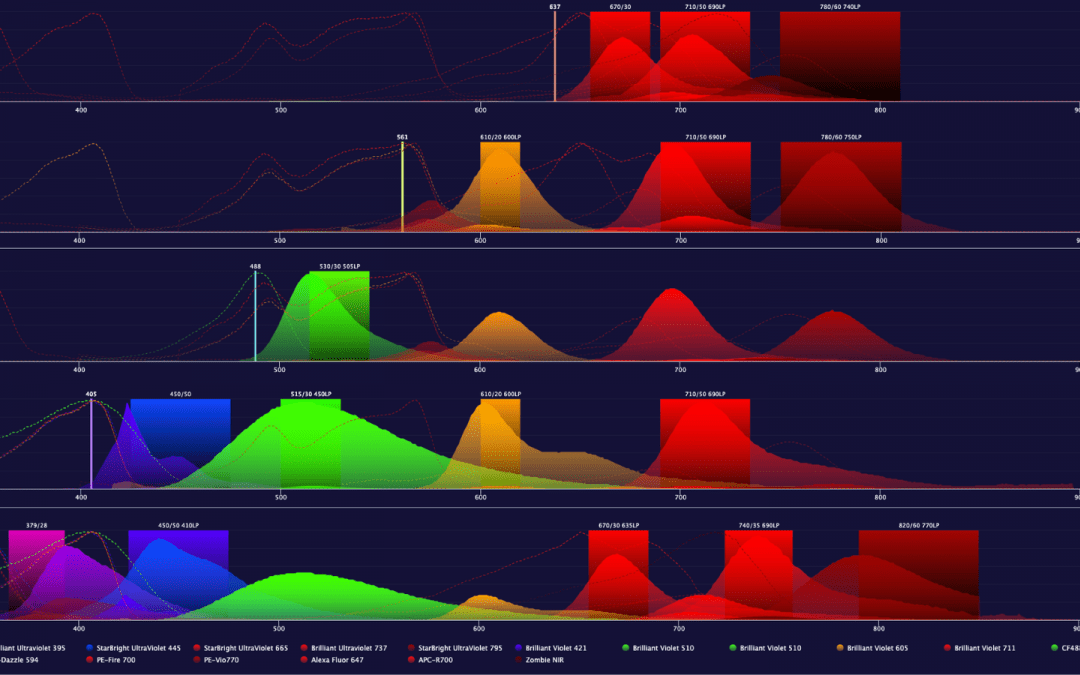
Flow cytometry is a technique for analyzing individual cells in suspension. It works by using a stream of fluid to direct the cells in single file past an interrogation point, where one or more lasers are focused. As the cells cross the interrogation point, they disrupt the laser light, which is recorded as forward scatter (FSC) and side scatter (SSC) by detectors positioned in front of or adjacent to the laser source. By gating on FSC versus SSC, it is possible to classify major cell populations in blood samples or identify cellular debris. However, because more detailed sample characterization is usually required, most flow cytometry experiments involve using fluorophore-labeled antibodies to identify different cell types based on marker expression. Here, we look at some common challenges faced by researchers when performing flow cytometry experiments and suggest ways of overcoming them. For further troubleshooting tips, download our Flow Cytometry Guide.
Fluorescence Intensity is Too High
Unexpectedly high fluorescence intensity can most often be attributed to the instrument settings, the immunostaining protocol, or panel design. To address inappropriate instrument settings, users can try decreasing the laser power, reducing the gain, or optimizing the photomultiplier tube (PMT) voltages with the aid of suitable controls. Protocol adjustments to consider include changing the blocking step (e.g., switching to a different blocking solution or increasing the blocking time) and titrating antibody reagents to determine optimal concentrations. It is also recommended to take a close look at any washes – increasing the number of wash steps or including a low concentration of detergent in wash buffers will help to ensure that any unbound antibody reagents are removed. In terms of panel design, pairing dim fluorophores with abundant antigens is advised.
High Background
Like unexpectedly high fluorescence intensity, high background can result from inadequate blocking or washing, or a failure to optimize the concentration of antibody reagents. However, it can also be due to autofluorescence or the detection of non-specific cell types. Ways of tackling autofluorescence include ensuring that cells are not over-fixed, using fluorophores that emit in the red channel for detecting highly autofluorescent cell types (e.g., neutrophils), and minimizing the presence of dead cells. The latter can be achieved by keeping samples on ice, avoiding freeze-thawing, and including a viability dye in the staining panel for excluding dead cells during data analysis. To prevent non-specific cell types from being detected, it is suggested to use well-validated antibodies and to introduce an Fc blocking step into the immunostaining protocol.
Low Signal
Low signal may often be a consequence of antigen expression. If the target of interest is scarce, a bright fluorophore will be needed for its detection, whereas if the antigen is intracellular, the cells will require fixing and permeabilizing for antibodies to access their epitopes. Low signal may also be attributable to the antibody reagents. It is important to check that the antibodies being used are validated for the chosen sample type, species, and fixation method (if applicable), and to optimize antibody concentrations. In addition, low signal could be due to inappropriate fluorophore selection or handling. Fluorophores should be matched with the flow cytometer’s lasers and detectors, and must always be protected from light to prevent photobleaching.
Unexpected Cell Populations
Sometimes, when performing flow cytometry experiments, several distinct cell populations may be observed when just one population was expected. This could be due to multiple cell types expressing the same marker, which will require that the staining strategy be altered to accurately identify the population of interest. It could also result from antibodies binding non-specifically to dead cells, the presence of which can be minimized as described previously.
Unusual Scatter Properties
Unusual scatter properties typically arise from poor sample quality, with contamination or cellular damage being most often to blame. It is recommended that samples be handled with care, using proper aseptic technique, and that freeze-thawing and harsh vortexing or centrifugation be avoided. Data quality can also be enhanced by running samples through the flow cytometer as soon after staining as possible.
Abnormal Event Rates
When performing flow cytometry experiments, the event rate should be sufficiently high for results to be statistically relevant, but not so high that sample quality is compromised by an extended acquisition time. If the observed event rate is lower than that which was set, it could be due to the flow cytometer becoming clogged or the sample being prepared at an incorrect (too low) concentration. A higher than expected event rate could instead result from sample contamination, the presence of large quantities of cellular debris, or cell numbers being too high. Ways of addressing these issues include following best practice recommendations for sample preparation and using an automated cell counter instead of a hemocytometer for counting.
Supporting Flow Cytometry Research
FluoroFinder has developed a suite of tools to help you with panel design and avoid these challenges. FluoroFinder’s Panel Builder can walk you through the fluorophore selection process to ensure you’re using reagents optimal for your specific cytometer configuration and experimental biology. Our Panel Builder‘s antigen density feature can help you find fluorophores optimal for your target’s antigen expression level to help you avoid poor signals and high background noise. If you’re looking for a specific reagent, our Dye Directory is a useful resource to learn more about the spectral properties and applications of over 1,100 dyes and help you find products from all major suppliers.
Sign-up for our eNewsletter to receive regular updates about flow cytometry and other fluorescence-based techniques, including the very latest product offerings to reach the market.


