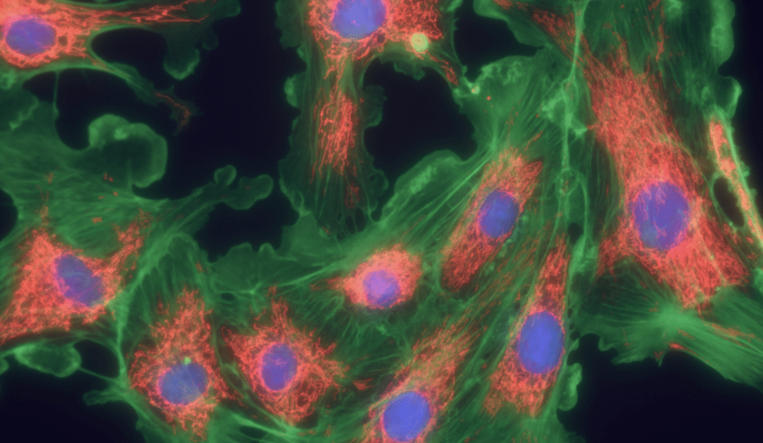
Intravital microscopy has vast potential to reveal hidden cellular mechanisms
Intravital microscopy (IVM) is a term used to describe the direct visualization of cells and tissues within a living organism. It was first reported in the 17th century, shortly after the invention of the microscope, but has since become increasingly sophisticated with the development of novel fluorophores and advanced microscopy techniques. Using modern methods, researchers can study complex physiological processes such as leukocyte extravasation into inflamed tissues, phagocytosis, and vascular development in real time, as well as investigate tumor growth and metastasis formation. Critically, being able to perform these types of experiments promises to reveal the cellular mechanisms underlying various physiological and pathological events.
Key Milestones
The road to intravital microscopy as we know it today began with Marcello Malpighi’s efforts in the mid-17th century to image the lungs of mammals and amphibians. A key milestone was reached in 1695, when Antonie van Leeuwenhoek improved microscope optics sufficiently to observe red blood cells in frogs, using a model which was subsequently adapted by Rudolf Wagner in the 1800s to study leukocyte rolling in the vasculature. In 1909, Julius Ries and Frédéric Vlès independently created the first movies of proliferating cells in the developing sea urchin. Then, in 1911, the invention of the fluorescence microscope marked a turning point for intravital microscopy by opening up new ways of observing biological specimens. The development of fluorescent antibody labeling by Coons et al. in 1942, and the cloning of the green fluorescent protein (GFP) by Chalfie et al. in 1994, provided further opportunities for more complex imaging studies. Methods that are now commonly used for IVM include confocal and laser scanning confocal microscopy (LSCM), multi-photon microscopy, second harmonic generation (SHG) microscopy, and fluorescence lifetime imaging microscopy (FLIM).
Intravital Microscopy Techniques
Although the earliest fluorescence microscopes were warmly received by the scientific community, they were soon discovered to suffer from high background due to scattered excitation illumination. This can be especially problematic for IVM, which typically uses thicker samples (that are more prone to scatter) than studies performed with cultured cells or tissue sections. Confocal microscopy was developed to address this issue, improving resolution by incorporating a pinhole to minimize interference from outside of the region of interest. A variation of confocal microscopy, known as laser scanning confocal microscopy, uses high-speed oscillating mirrors to pass a laser beam across the sample in a raster pattern, subsequently combining multiple focal planes to produce a 3D image. Confocal microscopy and LSCM tend to be used for IVM if the process being investigated occurs within 50–100 µm of the surface.
If the process of interest is located deeper than 50–100 µm below the surface, multi-photon microscopy is generally preferred. Multi-photon microscopy is based on the simultaneous absorption of two or more photons with wavelengths in the near infrared (NIR) or infrared (IR) region and allows for approximately five times the imaging depth of confocal microscopy methods1. Most multi-photon microscopes are fitted with a femtosecond pulsed laser that is focused to a single point, outside of which absorption is restricted, a feature which makes them more expensive than confocal instruments. The increased cost of multi-photon microscopes may, however, be outweighed by their superior sensitivity to weak fluorophores and reduced photobleaching compared to confocal microscopes, in addition to their increased penetration depth.
Another type of intravital microscopy technique, known as second harmonic generation (SHG) microscopy, involves the up-conversion of two low energy photons to produce a single photon that is exactly twice the frequency of the incident excitation source. It is used for imaging tissues that can generate a harmonic signal, predominantly those containing fibrillar collagen, which is found in the extracellular matrix (ECM) of connective tissues such as bone, cartilage, and tendons, as well as in the stroma of organs including the lung, liver, and kidney2. The imaging depth that can be achieved with SHG microscopy depends on the excitation wavelength but is broadly similar to that of multi-photon microscopy3. Third harmonic generation (THG) microscopy is a technique based on similar principles to SHG microscopy which can be used to complement this and other microscopy techniques.
Like THG microscopy, fluorescence lifetime imaging microscopy (FLIM) is often used alongside other microscopy techniques to provide deeper insights into the process of interest. With FLIM, the fluorescence lifetime of the fluorophore is measured (rather than its intensity) to provide a means of distinguishing fluorophores with identical emission wavelengths. Often, FLIM is used for resolving fluorophores that emit in the green spectrum (e.g., fluorescein) from tissue autofluorescence.
Intravital Microscopy Applications
A major advantage of intravital microscopy is that it provides information about cellular and tissue dynamics that simply cannot be obtained by other techniques. Specifically, intravital microscopy allows researchers to study complex processes as they occur within the native environment, without any need to artificially manipulate the underlying system. Of course, performing these types of experiments requires researchers to gain access to the tissue or organ being imaged. Transdermal and ocular imaging studies are the least invasive, often involving little more than affixing a coverslip to the tissue in question (if required by the objective lens) and immobilizing the region of interest. Accessing deeper tissues necessitates surgical exposure and, as such, these kinds of studies are almost always terminal for the animal involved. While the potential applications for intravital microscopy are virtually limitless, recent examples include the use of LSCM for monitoring hepatic lipid droplet accumulation in a murine model for nonalcoholic fatty liver disease; the imaging of cardiomyocytes in a beating murine heart with multi-photon microscopy; and the use of SHG microscopy for imaging contracting muscles in live Drosophila melanogaster larvae4,5,6.
Supporting Your Research
No matter which intravital microscopy technique you’re planning to use, FluoroFinder has the tools you need to help you design your experiment. Use our Fluorescent Dye Database to find detailed information on the optical properties and spectral profiles of over 1,000 fluorophores, then check out our Spectra Viewer to combine that information with instrument-specific laser and filter configurations.
Sign up for our eNewsletter to receive regular updates about a broad range of fluorescence-based techniques and discover the latest products available to streamline your research.


