Excluding dead cells from flow cytometry analysis with a cell viability fluorescent dye is critical for producing accurate results
The presence of dead cells in samples that will be analyzed by flow cytometry can affect antibody staining and lead to inaccurate conclusions. Although forward and side scatter can be used for gating out dead cells, this approach does not exclude all debris. For this reason, dyes capable of distinguishing live from dead cells are essential tools for those performing flow cytometry experiments. Viability dyes are categorized into three types – DNA binding dyes, protein binding dyes, and vital dyes – each offering different advantages and disadvantages. Researchers can minimize the presence of dead cells and debris by incorporating a cell viability fluorescent dye into their flow cytometry workflow to produce more reliable data and save time on repeating experiments hampered by false positive results.
Dead Cells and Debris Present Experimental Challenges
Whenever an experiment requires the use of cells, it is inevitable that there will be some level of cell death. Failure to determine the extent to which this has occurred can have far-reaching consequences depending on the application. During routine seeding, not accounting for the presence of dead cells can mean longer time taken to reach the desired confluence. For flow cytometry and cell sorting applications, non-specific antibody binding to dead cells reduces the dynamic range and makes it difficult to identify rare populations and weakly positive samples; this problem is exacerbated by the fact that dead cells exhibit high autofluorescence. In addition, dead cells release DNA, causing the formation of aggregates that can lead to the instrument becoming blocked.
Good Sample Preparation Technique Helps Minimize Cell Death
Cell death and unwanted aggregation can be reduced by following recommended practices for sample preparation. These include using freshly-isolated cells wherever possible, avoiding freeze-thaw cycles, and keeping samples at 4oC or on ice prior to use. It is also suggested that resuspension buffers be free of calcium (Ca2+) and magnesium (Mg2+) ions, and contain both EDTA and a low concentration of protein. Other recommendations include adding DNase to samples and passing cells through an appropriately sized strainer to remove any clumps before acquisition. The effectiveness of these measures can be confirmed by using a viability stain in the flow cytometry workflow.
A Cell Viability Fluorescent Dye Improves Flow Cytometry Data Accuracy
A wide selection of viability dyes, with a broad range of excitation and emission profiles, is available to researchers. Such dyes are grouped into three different categories, based on the mechanism of action. DNA binding dyes and protein binding dyes function by exploiting the compromised membrane integrity of dead cells, while vital dyes measure viability by fluorescing only when they are metabolized within living cells.
DNA binding dyes
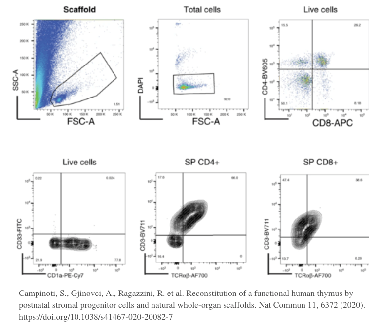
DNA binding dyes such as propidium iodide (PI), 7-Aminoactinomycin D (7-AAD), 4′,6-diamidino-2-phenylindole dihydrochloride (DAPI), and DRAQ7™ are the most commonly used viability dyes for flow cytometry. They are excluded from live cells with intact membranes, but are able to bind to DNA and fluoresce when the membrane has become leaky, providing a measure of cell death. Advantages of DNA binding dyes are that they are easy to use, simply requiring a brief incubation with the sample following antibody staining, and relatively inexpensive. However, DNA binding dyes are unsuitable for use with fixed samples, and should not be left on the cells for too long as they will stain every cell that dies during the course of the experiment. One way of accounting for this is to check the signal of one particular sample at both the beginning and end of the acquisition. Staining a sample of dead cells is also recommended as a compensation control.
Protein binding dyes
A major advantage of protein binding dyes such as Zombie Dyes™, Ghost Dyes™, Horizon™ Dyes, and Phantom Dyes is that they allow cells to be fixed after viability staining. Like DNA binding dyes, these reagents are membrane impermeant. However, they function by binding amine groups, which are sparse on the cell surface, but highly abundant inside the cell. Because amine binding produces a fluorescent signal, dead cells exhibit much higher fluorescence than their living counterparts. Protein binding dyes typically require a slightly longer staining time than DNA binding dyes and should always be used in the absence of free protein.
Vital dyes
Vital dyes fluoresce only when acted upon by living cells. Calcein acetomethoxyis (calcein AM) is the best known example, comprising a membrane permeable dye that does not fluoresce. When calcein AM enters metabolically active cells, cleavage of the acetomethoxyis group results in a fluorescent signal, meaning that living cells are identified by virtue of fluorescing bright green. Like DNA and protein binding dyes, vital dyes are quick and easy to use. Their main limitations are that they cannot be used with fixed samples and are more restrictive in the number of different products available.
Selecting the Right Cell Viability Fluorescent Dye
FluoroFinder offers a range of tools to help with selecting a suitable cell viability fluorescent dye for your flow cytometry experiment. Use our Spectra Viewer to quickly compare over 1,000 fluorophores from all suppliers in one intuitive platform, or turn to our Panel Builder to view the fluorophore and antibody offerings of >60 suppliers and design a panel in the context of your instruments and configurations. And, if you would like to discuss your experimental needs in more detail, our support team can offer expert guidance and support.
Sign-up for our eNewsletter to receive regular updates about flow cytometry and other fluorescence-based techniques, and be the first to hear about new viability stains coming on to the market.


