Flow cytometry allows researchers to characterize individual cells using fluorophore-labeled antibodies for detecting targets of interest. Sample preparation, antibody specificity, and panel design are all fundamental to producing reliable results. However, choosing the right flow cytometry buffers is equally as important. Flow cytometry buffers play a critical role not only in protecting epitopes and ensuring antibodies can access their targets, but also in preventing unwanted background signal that can lead to data misinterpretation. In this article, we share a few basic flow cytometry buffer recipes and highlight some of the commercial products developed to address common flow cytometry challenges.
Buffers Are Integral to the Flow Cytometry Workflow
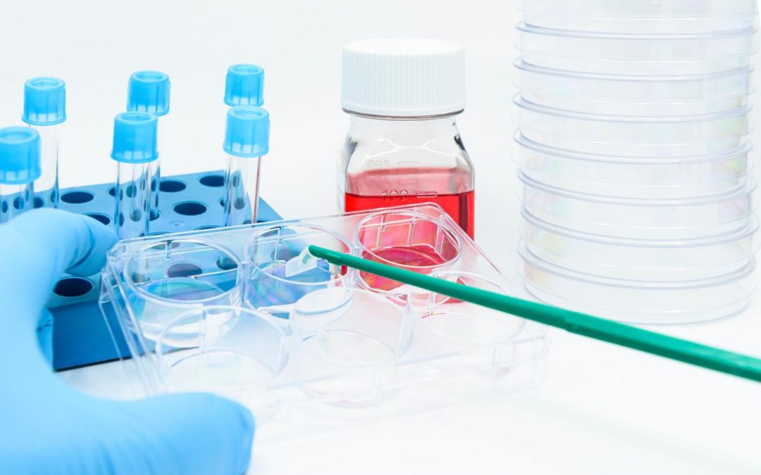
Cells routinely analyzed by flow cytometry, such as cultured cell lines, peripheral blood mononuclear cells (PBMCs), and tissue homogenates, must initially be processed to form a single-cell suspension. Then, the cells can be blocked and immunostained for surface markers or fixed and permeabilized before blocking and staining for intracellular targets. When detecting both intracellular and extracellular markers in the same experiment, it is customary to adopt a sequential staining protocol. This involves immunostaining for surface markers first to prevent damaging extracellular epitopes by fixation and permeabilization before detecting intracellular antigens. Buffers should be tailored to your model system. This involves optimizing the resuspension buffer, fixative, and permeabilization buffer, through to the blocking solution, antibody diluent, and wash buffer.
Resuspension Buffer
The resuspension buffer typically comprises of phosphate buffered saline (PBS) containing either 0.1-1% bovine serum albumin (BSA) or 1-10% fetal bovine serum (FBS) to reduce non-specific binding of antibodies and fluorophores to target cells. To prevent clumping, the PBS should be free of Ca2+/Mg2+. This buffer sometimes contains low concentrations of EDTA (0.5-5mM) and DNas and (25-50µg/mL) often includes 0.1-1% sodium azide (NaN3) as a preservative.
To avoid the variability associated with manually preparing resuspension buffers, some researchers prefer to use a preformulated solution. One option is BioLegend’s Cell Staining Buffer that also serves as an antibody diluent and wash buffer. “Our Cell Staining Buffer is optimized for immunofluorescent staining of viable or fixed single cell suspensions,” reports Kenta Yamamoto, Ph.D., BioLegend’s Product Manager for Cell Analysis. “Because it is free of biotin, calcium, magnesium, and EDTA, it is compatible with biotin/avidin indirect staining protocols.”
Red Blood Cell Lysis Buffer
Samples such as whole blood and tissue homogenates often contain residual red blood cells that prevent cells of interest from being collected in meaningful numbers. “For example, in peripheral blood there are so many red blood cells present that unless you collect millions of cells or increase the forward scatter (FSC) threshold you will only ‘see’ a few lymphocytes, monocytes and granulocytes,” explains Mike Blundell Ph.D., Global Product Manager at Bio-Rad. “The associated data files will be enormous, with only a few relevant cells in them.”
Because red blood cells can cause aggregation and are a source of autofluorescence, they should ideally be removed at the start of the flow cytometry workflow using a product such as Bio-Rad’s Erythrolyse Red Cell Lysing Buffer. “After incubating samples with Erythrolyse for 10 minutes at room temperature, the tubes are centrifuged and the supernatant decanted,” says Blundell. “The pelleted cells can then be washed and resuspended for processing with your usual protocol.”
Fc Blocking Buffer
While protein carriers prevent many non-specific binding interactions, flow cytometry protocols often require an additional Fc blocking step. “Immune cells such as monocytes, macrophages, natural killer (NK) cells, and B cells express Fc receptors at the cell surface, which can bind antibody reagents via the Fc domain,” says Blundell. “Our Human Seroblock and Mouse Seroblock are designed to prevent this type of interaction and ensure that only antigen specific binding is observed.” Fc blocking is performed prior to immunostaining with 5-10 minutes of incubation at room temperature.
Fixation and Permeabilization Buffers
Fixation typically involves first incubating the cell suspension in 1-4% paraformaldehyde diluted in PBS for 10-15 minutes at room temperature. The cells are then permeabilized with a further 10 minute incubation in a suitable buffer – usually 0.1-0.5% detergent (e.g., Tween®-20 or Triton® X-100) or 0.1% w/v saponin diluted in PBS. Because many permeabilization methods are reversible, it is important to include the permeabilizing agent in any antibody diluents and wash buffers that will be used downstream. In recent years, fixation and permeabilization buffers have evolved to streamline sample handling and better preserve targets of interest.
“Our Cyto-Fast™ Fix/Perm Buffer Set simplifies flow cytometry workflows by providing a single buffer set that contains reagents to allow cell fixation and permeabilization,” notes Yamamoto. “We have also developed True-Phos™ Perm Buffer for enhanced detection of intracellular phosphorylated targets, as well as other intracellular proteins such as Cyclin B1 and pan-STAT3.” Bio-Rad likewise offers products to improve detection of intracellular targets, including its Leucoperm reagent that is compatible with most commercially available monoclonal antibody conjugates.
Tandem Stabilizer
Tandem dyes consist of two covalently bound fluorophores, a donor and an acceptor. When the flow cytometer’s lasers excite the donor, photons are transferred to the acceptor via Förster Resonance Energy Transfer (FRET), resulting in the emission of light.
Tandem dyes increase flexibility in panel design by allowing researchers to obtain several readouts from a single laser. However, tandem dyes are commonly known for degradation or decoupling, which describes the loss of FRET to the acceptor. Degradation is often caused by light exposure, which can photobleach both the donor and acceptor molecules. Oxygen radicals can also damage acceptor dyes, complicating data analysis.
To prevent progressive tandem dye decoupling, BioLegend recently launched its Tandem Stabilizer, a novel buffer additive. This allows for storage of fixed, tandem-stained cells for up to four days prior to data acquisition, provided samples are kept at 4oC away from light.
Supporting Flow Cytometry Research
FluoroFinder has developed a suite of tools to aid flow cytometry based research. Before formulating your flow cytometry buffers, check out our Panel Builder to design your panel with fluorophores from all suppliers and visualize their spectral profiles in the context of your lab’s cytometer configuration. Then, optimize your buffer recipes to ensure your fluorescent signals are preserved.
Sign up for our eNewsletter to receive further updates about flow cytometry and other fluorescence-based techniques.


