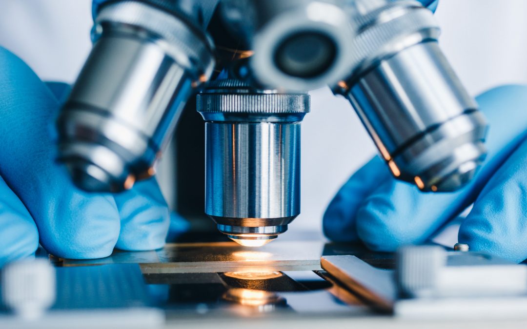
Microscopy is comprised of many innovative technologies and remains at the forefront of scientific research.
Since the invention of the microscope in the late 16th century, there has been a continual push to produce instruments with higher resolution, faster image capture, and the capacity to study living cells for longer with less detriment to their health. Advances in the field have been driven by the development of novel detection platforms, improvements to imaging and analysis software, and breakthroughs in sample preparation methodology, as well as by the production of countless modern fluorophores within more recent years. Unlike other techniques, microscopy allows researchers to localize and quantify sub-cellular structures and biomolecules to discover how sample composition and interactive processes vary under conditions of health and disease. Here, we provide an overview of some newer microscopy technologies and their defining features.
A potted history of microscopy
Hans and Zacharias Janssen are widely credited with inventing the microscope, which was first described in the 1590s. This early instrument consisted of a simple tube with lenses at either end and could magnify samples by up to approximately 20 times their normal size. During the course of the 17th century, the Janssen microscope was improved upon by scientists including Galileo Galilei, Anton Van Leeuwenhoek, and Robert Hooke, who introduced more sophisticated means of magnifying and focusing. Then came the invention of the ultramicroscope in 1903, the fluorescence microscope in 1911, and the electron microscope in 1938, before Gerd Binnig and Heinrich Rohrer developed the scanning tunneling microscope in 1981 to make 3D imaging possible. The field of microscopy continues to evolve and multiple technologies are now available to researchers, including the following examples.
Super-resolution microscopy
The term super-resolution microscopy (SRM) encompasses a range of technologies that were developed to overcome the 200 nm resolution limit of conventional light microscopy, which is imposed by the diffraction or scattering properties of light. These include methods such as Stimulated Emission Depletion Microscopy (STED) and Saturated Structured Illumination Microscopy (SSIM), which function by decreasing the size of the point spread function (the amount of blur associated with individual fluorophores). Other methods like Photo-Activated Localization Microscopy (PALM) and Stochastic Optical Reconstruction Microscopy (STORM), which sequentially visualize small numbers of photoswitchable fluorescent proteins or organic dyes. At present, SRM can achieve resolutions of as little as 10 nm and is being increasingly adopted across multiple research fields.
Expansion microscopy
Expansion microscopy breaks through the 200 nm diffraction barrier by using swellable polymers to physically expand the sample being investigated. Since expansion microscopy is compatible with standard microscopes and common fluorophores, this technique is growing in popularity, especially within the neuroscience community for synaptic imaging and neural circuit mapping in efforts to better understand neurological disease. Two main expansion microscopy workflows have been described: pre- and post-expansion labeling. The latter provides opportunities for signal amplification via methods including the hybridization chain reaction and iterative antibody binding.
Time-resolved electron microscopy
Time-resolved electron microscopy has its roots in scanning electron microscopy (SEM) and transmission electron microscopy (TEM), both of which use a pulse of electrons instead of light to create an image of the sample. Because electrons have much shorter wavelengths compared to light, SEM and TEM can magnify samples by up to 2 million times and 50 million times, respectively, achieving resolutions of ~0.5 nm (SEM) and <50 pm (TEM). The main difference between SEM/TEM and time-resolved electron microscopy is that the latter aims for short electron pulses at the sample, instead of a continuous electron beam, which enables temporal resolution. The pulses are typically delivered at femtosecond intervals and give rise to a series of snapshots offering detail of sample structure, composition, and – most importantly – dynamics.
Scanning helium microscopy
Scanning helium microscopy (SHeM) works by focusing a high-velocity beam of neutral helium atoms onto the sample, subsequently detecting any deflected atoms to produce a detailed image of the sample surface. One advantage of using helium is the reduced risk of sample damage as the neutral helium beams have lower energy compared to light or electrons. A next-generation form of SHeM, known as quantum gas jet-based helium atom microscopy (qHAM), is being developed to address the challenges associated with creating narrow beams of neutral particles to improve image quality.
Rapid Autofocus via pupil-split Image phase detection
Rapid Autofocus via Pupil-split Image phase Detection (RAPID) is a method compatible with light sheet microscopy which uses an auto-focusing technology for real-time correction of any artifacts introduced by the sample itself. First described in 2021, RAPID eliminates the need for tissue clearing (the use of chemicals to render tissues transparent), allowing for high resolution imaging of thicker samples that might otherwise introduce unwanted blurring. Using RAPID, researchers have been able to study whole mouse brains at the subcellular level, and have characterized both the 3D spatial clustering of somatostatin-positive neurons and the distribution of microglia.
Live-cell imaging microscopy
Live-cell imaging microscopy enables researchers to study cells without removing them from the controlled environment of the CO2 incubator. This is important to avoid cellular stresses that could lead to an altered phenotype or function. Included among the various live-cell imaging platforms on the market, CytoSMART Technologies’ Lux and Omni systems provide single-well or multi-well cell monitoring with both brightfield and fluorescent capabilities. Importantly, because the CytoSMART platforms work by moving the camera, not the culture vessel, they allow for the capture of highly detailed images without causing sample disturbance.
Supporting microscopy-based research
We offer a suite of user-friendly solutions to help you plan and execute microscopy-based research. Check out our Spectra Viewer with customizable microscope configurations to visualize over 1,000 fluorophores from all major suppliers or use FluoroFinder’s Antibody Search function to find antibodies for your targets of interest. Have a question? Our support team is always on hand to help guide you through experimental design.
Sign-up for our eNewsletter to receive further updates about microscopy and discover the latest trends within other research techniques.


