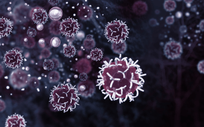
Articles
FluoroFinder News & Updates
From flow cytometry research and experimental design trends to FluoroFinder tool updates and industry applications, we explore it all in our blog.
Reagents for Super-Resolution Microscopy
Written by Sarah Locknar Reagents for Super-resolution microscopy Super-resolution microscopy (SRM) methods have taken the microscopy world by storm (literally!). These techniques enable imaging an order of magnitude beyond the optical “Raleigh limit”. There are two...
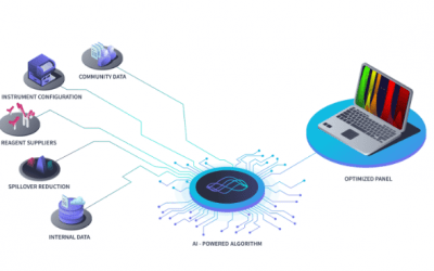
Simplify Flow Cytometry Panel Design with an All-in-One Platform
Written by FluoroFinder Staff Simplify Flow Cytometry Panel Design with an All-in-One Platform As flow cytometry technologies continue to advance, modern spectral and high-parameter platforms are enabling researchers to characterize complex cellular populations with...
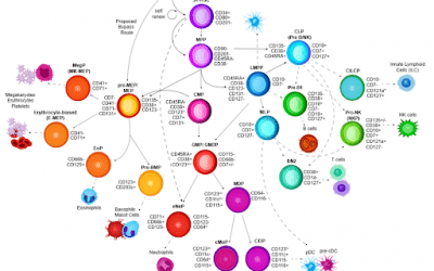
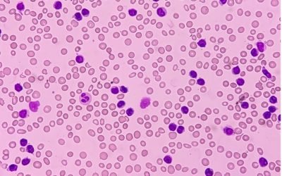
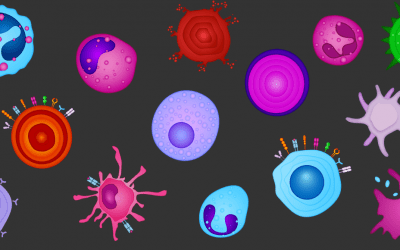
Cell Types and SOULCAP
Written by Kelly Lundsten Early in my career, I made the jump from neuroscience to immunology when I joined a biotech company that manufactured antibodies for flow cytometry. At the time, customers would send in lists of antigens and ask for help designing 17-color...
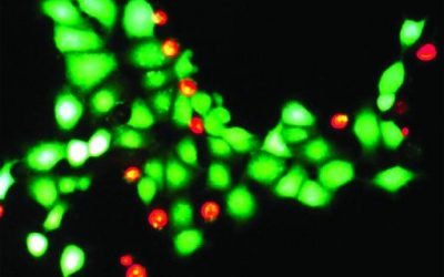
Flow Cytometry Viability Dyes: How to Measure Cell Health
Written by Emma Mason Flow Cytometry Viability Dyes: How to Measure Cell Health Dead and dying cells can significantly interfere with fluorescence-based techniques such as flow cytometry. Compared to healthy cells, non-viable cells often exhibit increased...
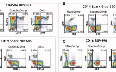
The Right Reference Controls for Spectral Unmixing
Sponsored by Slingshot, Written by Nathan Ni Spectral flow cytometry has proven revolutionary for studying and characterizing cells, giving scientists the power to investigate up to fifty different fluorophores in a single panel. This lets them delve deeper into...
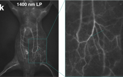
Invisible Imaging: Fluorescence Microscopy in the UV and NIR
Written by Sarah Locknar The whole-body fluorescence imaging of a mouse IV-injected with the NIR-II dye IDSe-IC2F encapsulated via PEGylation, excited with 793 nm laser light. Used under CC by 4 license without modification from (Feng, 2021) Figure 8k Invisible...
Flow Cytometry Experiment Design: Tools for Panel Optimization
Written by Sarah Locknar Tools for Flow Cytometry Experiment Design Designing a successful flow cytometry experiment requires considerable effort and skill. Not only must you understand the expected location and expression of different cellular markers, but you must...
Clinical Flow Cytometry: Where Measurement Meets Medicine
Written by Paul E. Mead, BSc (Hons), PhD, SCYM (ASCP) Clinical Flow Cytometry: Where Measurement Meets Medicine From Bethesda consensus to high-dimensional cytometry and the next era of clinical diagnostics Many consequential decisions in medicine begin with a...
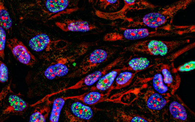
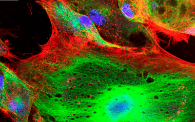
IHC vs. ICC vs. IF: What’s the Difference?
Written by Sarah Locknar IHC vs ICC vs IF: Differences in Immunolabeling Techniques What Are IHC, ICC, and IF? Immunohistochemistry (IHC), immunocytochemistry (ICC), and immunofluorescence (IF) are closely related techniques that use antibodies to detect antigens in...
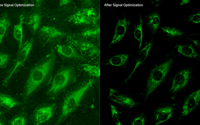
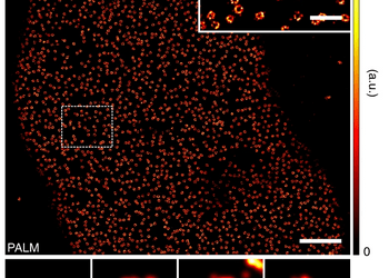
Increasing signal to noise ratio in fluorescence microscopy and blocking methods
Written by Sarah Locknar Blocking and signal-to-noise enhancement in immunofluorescence imaging Advanced fluorescence microscopy techniques like TIRF, STORM and PALM, depend on high signal-to-noise ratios (SNR) to detect small signals down to single-molecules. In this...
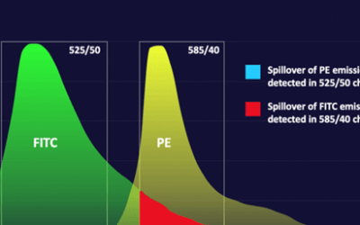
Flow Cytometry Controls: Essential Types, Setup, and Best Practices
Written by Sarah Locknar Flow cytometry controls serve several functions. They standardize and verify instrument performance, compensate for spectral spillover, verify specificity and sensitivity of staining, and help set gates for detection. Some controls are...
Fluorophores 101: Types, Brightness, and How to Choose the Right Dye
Written by Kelly Lundsten It can be a challenge to navigate the reagent catalogs of vendors in the life sciences. New brand names are popping up all the time and it is difficult to know if they are truly emerging technologies or if they are simply the same chemistries...
How to Use Cell Sorting for Single-Cell RNA Sequencing (scRNA-seq)
Written By Rea Dabelic Single-cell RNA sequencing (scRNA-seq) has transformed how researchers study cellular heterogeneity in complex tissues. However, the quality of a single-cell experiment depends heavily on sample preparation and cell isolation, particularly when...
Fluorescence Imaging Instruments: Microscopes, Plate Readers, and Spatial Platforms
Written by Emma Mason Instruments for fluorescence imaging allow researchers to study a vast array of biological processes, typically aided by labels in the form of protein tags or synthetic dyes. Fluorescence imaging instruments range from gel documentation systems...
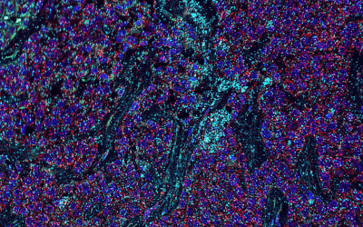
Multimodal and Multiplexed Spatial Imaging: A Practical Guide
Cover Image: Multiphoton (red) and Second Harmonic Generation (green) image of a human carotid cross-section. Used without modification under Creative Commons Attribution 4.0 International License from (Marchetti, 2019). Written by Sarah Locknar, Ph.D. From imaging...
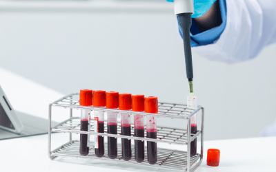
Flow Cytometry Sample Preparation: Tips, Protocol Steps, and Best Practices
Written by Emma Mason When Benjamin Franklin coined the phrase “by failing to prepare, you are preparing to fail”, he certainly wasn’t referring to flow cytometry, which wouldn’t be invented for another 200+ years. However, the expression is apt—good sample prep is...
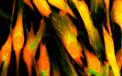
Live-Cell Migration, Invasion, and Adhesion Assays: Methods and Imaging Approaches
Written by Emma Mason Cell migration is fundamental to a broad range of physiological processes, including embryological development, angiogenesis, tissue regeneration, wound healing, and immune responses, to name just a few. It also has a pivotal role in the...

A Look Back on Flow Cytometry in 2025
Guest Authored By: Kelly Lundsten There aren’t many people left who need to be convinced that spectral cytometry, the pivotal coalescence of engineering and computational advancements that has enabled 401-5 and 506 color flow cytometry, is destined to become a...

New Dyes of 2025 & Expansion of Spectral Dyes
Every year, the number of fluorescent dyes available for scientific research continues to grow, and 2025 has been no exception. Novel fluorochromes are expanding the limits of both traditional and spectral flow cytometry, as well as advancing spatial biology...
Imaging Flow Cytometry
For decades, traditional flow cytometry has been the dominant technique for analyzing single cells within large, heterogeneous populations. However, imaging flow cytometry (IFC) is rapidly becoming more mainstream due to the advantages that it can provide. We spoke...
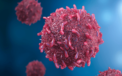
Advancing Cytometry Technology in Oncology
Guest Authored By: Kelly Lundsten I often think about a concept called the pinball effect. Distilled, it is the knowledge of all of the small changes made by people whose names are remembered, or not, who optimized a process or invented a new chemistry or asked “why”...
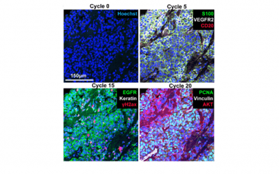
Automation & Microfluidics in Cyclic Immunofluorescence
Cover Image: Selected images of a melanoma specimen subjected to 20 cycles of Cyclic IF after cycles 0, 5, 15 and 20. Used without modification under Creative Commons Attribution 4.0 International License from (Lin, 2018) Figure 4G. Spatial biology aims to...
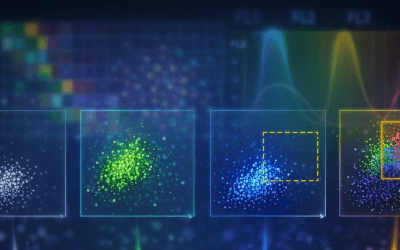
New and Emerging Spectral Flow Cytometry Instrumentation
Spectral flow cytometry improves on traditional flow cytometry by capturing the entire emission spectrum of each fluorochrome, rather than isolating specific wavelengths. This enables more precise resolution of overlapping fluorochromes and greater flexibility in...
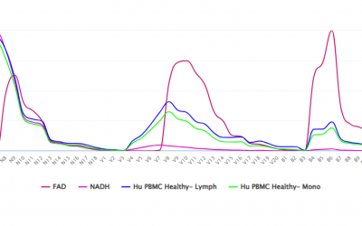
Autofluorescence in Flow Cytometry
Guest Authored By: Kelly Lundsten Every living organism on earth has evolved to either harness or protect itself from the energy of the sun. Organic molecules capture energy and conduct it along a circuitry of electron pairs shared by double bonds that compose various...
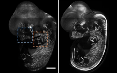
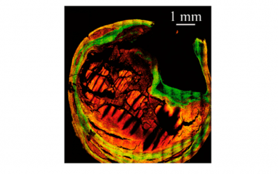
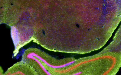
Clearing Tissue for Microscopy: Part 2 – Hydrophilic Methods
Cover Image: E12.5 whole mouse embryo was immunostained with neurofilament antibody and cleared with RTF. Scale bar, 1000 μm. Used without modification under Creative Commons Attribution 4.0 International License from (Yu T. Z., 2018) Figure 4a. As explained in last...
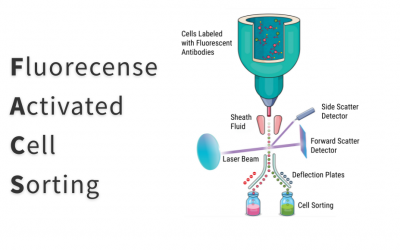
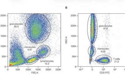
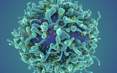
All About FACS
Fluorescence-activated cell sorting (FACS) is an advanced form of flow cytometry that allows researchers to isolate specific cell types from a heterogeneous sample. This article explains the basic principles of FACS and describes some common research applications. It...
Clearing Tissue for Microscopy: Part 1 – Dehydration Protocols
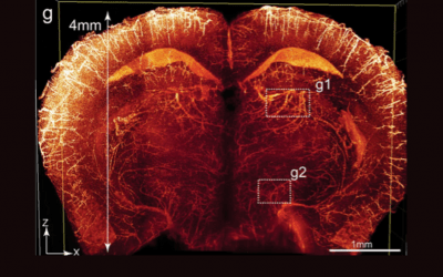
Cover Image: Imaging of the whole-brain vasculatures at sub-cellular resolution. Brains of adult Tie2-Cre;tTAflox;tetO-H2BGFP (TTH) mice were cleared using the PEGASOS method and imaged with a tiling light-sheet microscope. g An X-Z optical slice acquired at the...
Blockers, Buffers, and Beyond: Supplementary Reagents for Flow Cytometry
When selecting reagents for flow cytometry, fluorescently-labeled antibodies are likely the first products that come to mind. However, supplementary reagents are also important and it pays to investigate what’s on offer. This article looks at some of the different...
Flow Cytometry of EVs and Other Small Things
Guest Authored By: Vera Tang, Ph.D. Interest in the study of extracellular vesicles has increased exponentially over the past two decades due to the important role they play in inter-cellular communication, as biomarkers of disease, and vehicles for drug delivery....
Fluorophore Families
So many new fluorophores have been released in recent years, it can be hard to keep track of their similarities or novelty. Sometimes, a company will create a new brand name for a fluorescent chemistry that is already commonly used, which can make sifting through the...
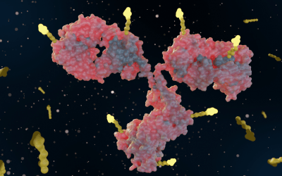
Antibody Conjugation Techniques
Conjugating antibodies to other molecules is a common technique used for drug delivery, changing solubility, attaching to solid substrates (for purification or assays), and visualizing the antibody using fluorescent, magnetic or gold nanoparticles. Western blots,...
Cell Mimics In Cell Manufacturing Applications
Flow cytometry assays are integral to cell manufacturing, where they have an essential role in safeguarding product quality and function. However, the types of controls used for flow cytometric characterization are changing as researchers realize the advantages of...
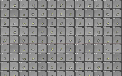
A Look at High-Content Imaging
High-content imaging (HCI) combines automated fluorescence microscopy with advanced image analysis to study cellular samples. This article looks at the key steps in a typical HCI workflow and explores some common applications. It also highlights some of the leading...
Advantages of Recombinant Antibody Development
Recombinant antibody technology is addressing known limitations of traditional antibody molecules and opening up new areas of scientific research. We spoke with Michael Fiebig, Ph.D., Chief Scientific Officer at Absolute Antibody (a Vector Laboratories Company), Mary...
The Rise of Spatial-Omics Technologies
Single-cell analysis techniques such as fluorescence-activated cell sorting (FACS) and single-cell DNA and RNA sequencing have broadened researchers’ understanding of cell biology in health and disease by enabling the study of rare cell types and phenotype variations...
Latest Developments in Flow Cytometry
Flow cytometry continues to become more powerful with the development of novel instrumentation and reagents. Here, we highlight some of the latest advances in the field and look at how they can benefit your research. Thermo Fisher Scientific Launches the Invitrogen™...
The Discovery and Evolution of Fluorescent Proteins
It has been over 60 years since Osamu Shimomura et al. discovered Green Fluorescent Protein (GFP)1. Since then, the color palette for fluorescent proteins has been extended to span blue through to far red, and even includes proteins that are capable of exhibiting...
Steric Effects in Multiplexed Immunofluorescence
Structural biology is focused on mapping where proteins, lipids and other biological molecules are located in cells and tissues in space. Researchers often use immunofluorescence labelling techniques that have been around for decades to visualize biomolecules and...
Definitive Phenotypes in Flow Cytometry
Standardization may not be the most exciting topic in biomedical research, but in an era where we lament the lack of reproducibility and distribute blame to reagents, sample prep, and general technical know-how, standardization is something every researcher should...
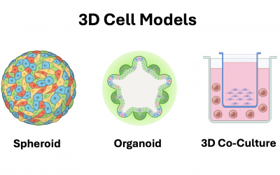
2D vs 3D Cell Cultures
Two-dimensional (2D) cell cultures represent a cornerstone of scientific research due to their relative simplicity and the establishment of standard techniques over time. In recent years though, 3D cell culture models have risen in popularity for the more...