Articles
FluoroFinder News & Updates
From flow cytometry research and experimental design trends to FluoroFinder tool updates and industry applications, we explore it all in our blog.
Fantastic Beasts and How To Image Them
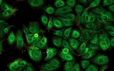
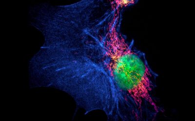
Fluorescence microscopy is seeing increased utility for imaging organisms While fluorescence microscopy has long been employed for imaging plated mammalian cells, its use for visualizing organisms is flourishing. In recent years, winning images in Nikon’s annual Small...
Preparing a Single-Cell Suspension for Flow Cytometry
High viabilities, well-preserved antigens, and minimal clumping underpin flow cytometry success Unlike bulk population analysis techniques such as ELISA and Western blot, flow cytometry provides information about individual cells. Although microscopy does this as...
OMIPs
What are OMIPs? Optimized multicolor immunofluorescence panels, commonly known as OMIPs, are highly validated sets of antibodies and reagents that are published, peer-reviewed, and compiled to serve as a resource to the scientific community. OMIP publications often...
Advances in Western Blot Technologies
New approaches to Western blotting techniques overcome performance limitations For over 40 years, researchers have used Western blot for protein identification. Not only does Western Blot provide confirmation that a target of interest is present in a sample, this...
Expansion Microscopy
Expansion Microscopy physically expands biological specimens enabling higher resolution imaging First described in 2015, expansion microscopy is an imaging technique that improves on the resolution of conventional light microscopy by physically expanding the...
Flow Cytometry in Stem Cell Research and Therapy
Identifying distinct stem cell types and monitoring their differentiation is essential for developing novel therapies Stem cell research originated in the 1950s when it was discovered that mice which had received a lethal dose of radiation could recover...
Excluding Dead Cells With a Cell Viability Fluorescent Dye
Excluding dead cells from flow cytometry analysis with a cell viability fluorescent dye is critical for producing accurate results The presence of dead cells in samples that will be analyzed by flow cytometry can affect antibody staining and lead to inaccurate...
Newsletter: Imaging and Analysis of 3D Cell Cultures
Compared to traditional monolayers, 3D cell cultures more closely resemble physiological conditions 2-dimensional (2D) cell cultures consisting of monolayers attached to a flat surface have been a mainstay of scientific research for decades. However, cells grown in...
Newsletter: Considerations for Spectral Flow Cytometry Panel Design
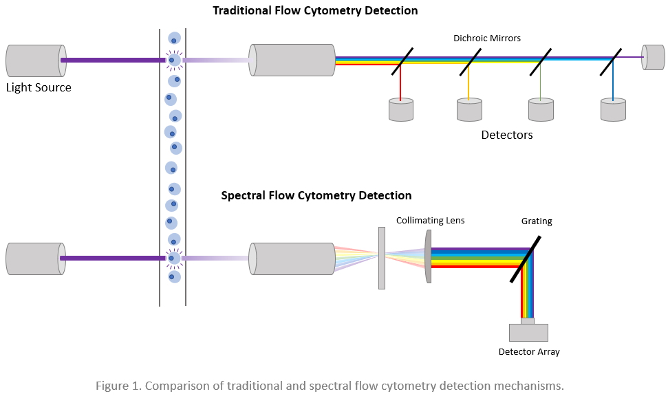
Spectral flow cytometry offers several advantages over conventional flow cytometry but may be more affected by spreading error Spectral flow cytometry was first described in 2004 by the Robinson group at Purdue University Cytometry Laboratories. Since then, several...
Newsletter: Confocal Microscopy and Related Technologies
Confocal microscopy offers several advantages over traditional widefield microscopy and has evolved to meet many different research needs Originally developed by Marvin Minsky in the 1950s to address the problem of light scatter, confocal microscopy has become an...
Newsletter: Distinguishing Cell-Cell Complexes from Singlet Cells
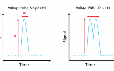
Failure to correctly identify cell-cell complexes can lead to data being misinterpreted and compromise the purity of sorted cell populations Multiparametric flow cytometry allows researchers to detect different cell types within a heterogeneous population. When...
Newsletter: Antibody Selection Guidelines
Choosing the right antibody for the intended application and sample type is critical to producing reliable results Antibodies are essential tools for applications ranging from flow cytometry, immunocytochemistry (ICC), and immunohistochemistry (IHC) to Western...
Newsletter: Comparison of Flow Cytometry Analysis Software
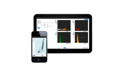
Software packages for viewing and analyzing FCS data files offer different features and benefits Data analysis represents a major source of variation in flow cytometry. Not only is the analysis process highly prone to user subjectivity, but disparities may arise due...
Newsletter: Fluorescent proteins: advantages and disadvantages
Fluorescent proteins have enabled countless scientific discoveries, but they have their limitations For decades, researchers have been fascinated by the ability of certain living organisms to emit light. However, it was not until 1962 that this phenomenon was first...
Newsletter: CRISPR as an Imaging Tool
Repurposing the CRISPR-Cas system for imaging genomic targets in living cells Established methods for cell imaging use fluorophore-labeled antibodies or oligonucleotides to bind proteins or nucleic acids in fixed cells or rely on the engineered expression of...
Introduction to Spectral Overlap and Compensation in Flow Cytometry
Compensation is one of the most important but least understood aspects of multicolor flow cytometry Multicolor flow cytometry allows researchers to characterize complex cellular populations by staining for several cell type specific markers at the same time. However,...
Newsletter: Tyramide Signal Amplification in Microscopy and Spatial Proteomics
Advantages of TSA include increased sensitivity, simplified panel design, and faster time to results Tyramide signal amplification (TSA), also known as catalyzed reporter deposition (CARD) or tyramine amplification technique (TAT), is a method used for detecting...
Newsletter: Past, Present, and Future of Flow Cytometry
Flow cytometry continues to evolve with advances in instrumentation, reagents, and software The first fluorescence-based flow cytometer was developed in 1968 and was commercialized the following year. Since then, the field of flow cytometry has matured rapidly, and...
Newsletter: Fluorescent Probes for Intracellular Calcium Measurement
Studying calcium flux leads to insights into normal physiology and disease Calcium ions (Ca2+) are involved in numerous intracellular signaling cascades and in a broad range of physiological processes. These include neurotransmission, muscle contraction, and hormone...
Newsletter: Cellular Markers for Flow Cytometry Analysis
Marker selection and panel design underpin the accuracy of cellular identification Being able to monitor the presence and relative abundance of different cell types in sample material is essential to understand normal biology and investigate the pathogenesis of...
Newsletter: Dyes for Analyzing the Cell Cycle and Apoptosis
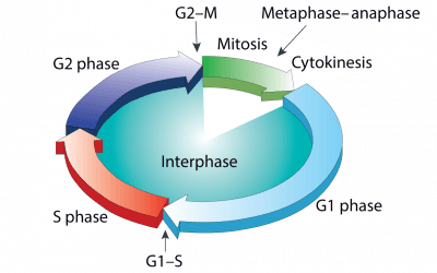
Aberrant cell proliferation and cell death underlie a multitude of disease states Normal tissue homeostasis depends on a critical balance between cell proliferation and cell death. The cell cycle regulates the former, while the latter occurs via controlled...
Newsletter: Fluorophores for Confocal Microscopy
Confocal microscopy is a powerful imaging technique used to study biological specimens. It offers several advantages over conventional widefield microscopy, including the capacity to control depth-of-field and collect serial sections from thick samples. Fluorophores...
Newsletter: Flow Cytometry Applied to Drug Discovery: Critical Reagent Monitoring
Best practices for antibody and fluorophore use safeguard assay performance A defining feature of flow cytometry is its capacity to analyze single cells. This has led to its application across the entire drug development continuum, with recent advances in the field of...
Newsletter: Designing a Fluorescence Microscopy Experiment
For over 100 years, researchers have used fluorescence microscopy to study biological samples like plated cells or tissue sections. Such experiments typically involve using fluorophore-labeled antibodies to recognize and bind targets of interest before imaging with a...
Newsletter: Analysis of Rare Cell Populations
Rare cell types provide novel insights into mechanisms of health and disease Many techniques used for cell-based research rely on bulk analysis, with data being generated from a complex heterogeneous population. The main drawback of these methods is that they can lead...
Newsletter: Overview of Advanced Fluorescence Microscopy Techniques
Conventional fluorescence microscopy uses fluorophore-labeled antibodies to visualize cellular structures, providing insights into cellular physiological and pathological states. However, the wave nature of light imposes a resolution limit of approximately 200 nm on...
Newsletter: Single-Cell Analysis in Immunology
In recent years, technologies for single-cell analysis have evolved rapidly, revealing huge differences between cells once categorized as being the same type. This variability is most apparent within the cells of the immune system, which must undergo multiple dynamic...
Newsletter: OMIP Resource and Validated Panel
The “optimized multicolor immunofluorescence panel” OMIP publication format was launched 11 years ago as a collaborative platform to establish criteria for experimental design, data collection, and analysis [1]. An OMIP is, by definition, a peer-reviewed publication...
Newsletter: Imaging Flow Cytometry
A technique that combines flow cytometry with digital microscopy promises novel insights Imaging flow cytometry (IFC) has seen increased uptake in recent years, largely due to instrumentation and software improvements that have made the technique more accessible. By...
Newsletter: History of fluorescent dyes
The widespread adoption of fluorescence applications is a testament to the importance of fluorescent dyes in biochemical and biological research. The speed, reproducibility, and sensitivity of fluorescence-based analysis methods explain their rapid adoption in all...
Newsletter: Essential Flow Cytometry Bookmarks
Typing “flow cytometry” into the Google search box returns 11,300,000 results! A daunting amount of information to process for anyone approaching the technology for the first time. To help anyone wanting to learn more about flow cytometry navigate this huge body of...
Newsletter: Spectral Flow Cytometry
The recent publication of a 43 color panel for the characterization of Peripheral Blood Mononuclear Cells (PBMC) is yet another example of the multiplexing potential of spectral flow cytometry. The technology, first demonstrated by Dr. Paul Robinson at Purdue...
Newsletter: Reducing Background Signal with Recombinant Antibodies
Enhance the quality of your flow cytometry data by using recombinant antibodies Recombinant antibodies have many advantages over conventional antibody reagents. These include exceptional batch-to-batch reproducibility, highly scalable in vitro production, and the...
Newsletter: Fluorophores for Super-Resolution Microscopy
Breaking through the diffraction barrier for deeper biological insights Super-resolution microscopy (SRM) improves conventional fluorescence microscopy by achieving higher resolution. It enables fine mapping of structures such as microtubule protofilaments,...
Newsletter: Gating Strategies
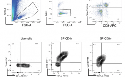
Analysis of flow cytometry data requires the identification of a homogenous cell population. This is achieved in a multistep process known as gating. It involves narrowing down the parameters that define the cell population of interest and excluding dead cells and...
Newsletter: From Research to Therapy: How Flow Cytometry Supports the Drug Discovery Pipeline
From Research to Therapy Flow cytometry is widely used, with many applications in both research and clinical labs. For pharma and biotech labs, the ability to quickly perform multi-parameter quantitative analysis on large numbers of heterogeneous cells makes flow...
Newsletter: Dyes for Spectral Analyzers
Maximize the benefits of spectral flow cytometry by using the right fluorochromes Spectral flow cytometry enables researchers to conduct larger and more comprehensive flow cytometry experiments than ever before, in some cases with over 40 different fluorochromes in a...
Newsletter: New Technology Roundup
Some of the latest developments advancing scientific research With 2021 now well underway, we felt it might be timely to highlight some of the newer technologies available to enhance laboratory workflows. Modern dyes designed to increase panel size have proven hugely...
Newsletter: Flow Cytometry Analysis of Primary Cell Types: Moving Beyond Immortalized Cell Lines
Moving Beyond Immortalized Cell Lines For decades, immortal cell lines such as HeLa, HEK 293, A549, and Jurkat have provided researchers a useful and inexpensive platform in which to model normal and disease cellular pathways. However, researchers are increasingly...
Newsletter: Tips and Tools for Flow Cytometry Sample Prep Success
Improve your flow cytometry sample prep for more reliable results Sample material is precious, so it’s essential that flow cytometry data is both reliable and reproducible. This means optimizing every step of your flow cytometry protocol, beginning with sample prep,...

