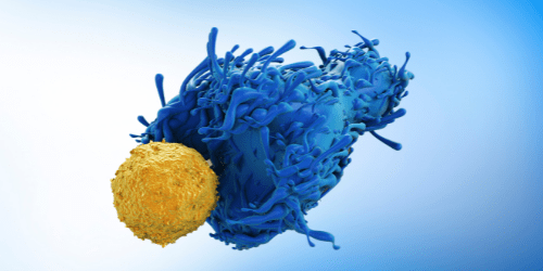
Rare cell types provide novel insights into mechanisms of health and disease
Many techniques used for cell-based research rely on bulk analysis, with data being generated from a complex heterogeneous population. The main drawback of these methods is that they can lead to rare events being missed due to masking by more prevalent cell types. Flow cytometry addresses this problem by allowing multiple measurements to be performed on a cell-by-cell basis. Yet, using flow cytometry to detect and analyze rare cell types presents inherent challenges. In this article we explain what constitutes a rare cell population, providing some examples, and comment on why rare cell types can be so difficult to analyze. We also suggest some strategies to enhance rare cell detection.
What is a rare cell population?
Broadly speaking, a cell is considered to be rare when it represents <0.01% of the total population. Examples of rare cell types include circulating tumor cells present in the blood of patients with solid tumors, which can be used to monitor disease and predict clinical outcomes, and circulating fetal cells found in the blood of pregnant women that enable non-invasive prenatal testing. Other rare cell populations include antigen-specific lymphocytes that may, for example, be used to study the immune response and develop effective immunotherapies and hematopoietic stem cells that offer significant potential for a broad range of tissue engineering applications.
Challenges of rare cell population analysis
One of the biggest hurdles that must be overcome when analyzing rare cell populations is collecting enough events to obtain statistically significant data. In flow cytometry, an event is defined as a voltage pulse correlating to the detection of a single particle by the instrument and may result from either cells or debris. When analyzing rare cell populations, it can often be necessary to collect millions of events for statistical robustness; the exact number will be dictated by the abundance of the cell type of interest in the sample, the signal-to-noise ratio of the detected cells over background fluorescence, and the percentage of cells present compared to debris. A further challenge arises from the fact that multiple markers are typically needed to accurately detect the target cell population; for example, to identify a specific lineage subset, activation state, or memory status, as many as ten or more markers may be required, increasing the complexity of panel design.
Acoustic focusing flow cytometry increases throughput
Acoustic focusing flow cytometry was developed to address the need for higher acquisition rates (translating to shorter experimental workflows), a key requirement for rare cell population analysis where data accuracy depends on collecting large event numbers. Unlike conventional hydrodynamic flow cytometry that aligns cells using hydrodynamic forces, acoustic focusing flow cytometry uses ultrasonic waves to focus cells for laser interrogation. This allows for higher sample input, increasing throughput and data acquisition speeds, and also results in less sample clogging. A further advantage of acoustic focusing flow cytometry is that it supports the use of large sample volumes; this can be beneficial when working with dilute material such as cerebrospinal fluid.
Innovative tools enhance specificity and sensitivity
Advances in flow cytometry instrumentation and the development of novel fluorophores spanning the entire visible to near-infrared spectrum have dramatically improved the specificity of rare cell detection. Now, by employing gating strategies based on sophisticated multiparameter analysis, researchers can combine a greater number of markers to both identify the population of interest and eliminate unwanted cell types from the analysis. In turn, using more markers increases assay sensitivity, which has additionally been enhanced by the development of sample preparation methods designed to better preserve cellular integrity. Examples include Thermo Fisher Scientific’s High-Yield Lyze for eliminating red cells from whole blood with minimal loss of rare blood cell populations and BD Bioscience’ Horizon™ Dri Tumor & Tissue Dissociation Reagent (TTDR) that helps maximize cell yields for single-cell studies while minimizing cell death and epitope damage. Reagents such as Luminex Corporation’s Muse® Count & Viability and Miltenyi Biotec’s Viobility™ Fixable Dyes are widely used to assess the impact of sample preparation methods on flow cytometry results.
Pre-enrichment improves detection
By pre-enriching specific cellular subsets before performing flow cytometric analysis, the frequency of rare cell types can be increased to improve detection rates. Strategies for pre-enrichment include methods that exploit physical properties of cells – for example, a hypotonic medium can be used to lyse red blood cells, leaving behind nucleated cells that are more resistant to hypotonic shock over time – and techniques based on immunological characteristics. A popular example of the latter is the use of antibody-conjugated magnetic beads to extract target cell populations from solution through bead binding to cell surface markers; numerous publications cite this approach and it has been employed for enrichment of circulating tumor cells, antigen-specific T cells, antigen-specific B cells, and hematopoietic stem cells, to name just a few.
Reagent selection for rare cell population analysis
Panel design is critical to the success of rare cell population analysis, where multiple markers must often be combined for accurate identification of target cell types. FluoroFinder’s online tools streamline panel design, freeing up researchers’ time to be spent on other tasks. Our Spectra Viewer lets you quickly compare over 1000 fluorophores from all suppliers in one intuitive platform, many of which have been literature-cited for rare cell detection, while our Panel Builder helps you optimize your multiplexed experiments with the very latest fluorophore and antibody offerings across >60 suppliers. If you need further advice, just contact our technical support team – we’re always happy to help, whatever the aim of your project.


