
The “optimized multicolor immunofluorescence panel” OMIP publication format was launched 11 years ago as a collaborative platform to establish criteria for experimental design, data collection, and analysis [1]. An OMIP is, by definition, a peer-reviewed publication reporting on carefully validated antibodies, reagents, and protocols for the multiparameter characterization of a specific cell state or response. Together with the MIFlowCyt standard (minimal information about a flow cytometry experiment) [2] initiative, it addresses the need to generate consistently reproducible data. It is also a way to acknowledge the efforts of the developer of the panel through the citation of the publication.
What is included in an OMIP?
OMIPs are published in the journal Cytometry Part A (Wiley Online Library) and include all the necessary information required for the execution of the panel, including:
- Purpose specific uses and limitations of the panel.
- A detailed list of antibodies, fluorochromes, and reagents.
- Panel development and optimization strategy.
- Staining method.
- Information on antibody titration.
- Controls.
- FCS data files.
- Gating Strategy.
To date, the majority of the OMIPs published have been developed for flow cytometry, however, the original intent was for this type of communication to be applied to any multiparameter fluorescence-based method.
OMIPs’ Numbers
81 OMIPs have been published since 2010. The rate of OMIPs generation has been steadily growing since, as a testament to the validity of this type of publication. OMIPs are addressing the demand of the scientific community for a transparent and efficient way to share information. OMIPs publications have been cited more than 500 times; OMIP-001, 003, 007, 014, 017, and 044 are the most cited. OMIPs panel size has also been growing, reflecting both the need to extract as much information as possible from a cell population and the increased multiplexing potential brought about by technological innovations [3] [4].
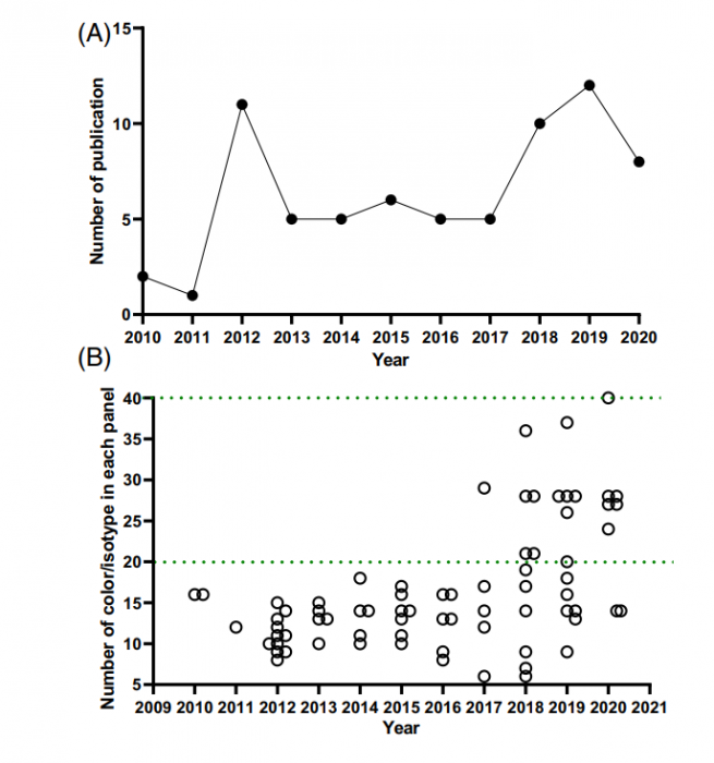
OMIP yearly number of publications (A) and panel size (B) [4]
COMING SOON: Access all OMIPs on Finder’s Flow cytometry Panel Builder
All existing OMIPs will be integrated into our newly updated panel builder. In just a few clicks you will be able to use FluoroFinder’s panel design tool to upload, modify and tailor OMIPs to your experimental needs, or to design an entirely new panel based on an existing one.
References
- Roederer M, Tarnok A. OMIPs—orchestrating multiplexity in polychromatic science. Cytometry A. 2010;77:811–2. 3 https://pubmed.ncbi.nlm.nih.gov/20722007/
- Lee JA, Spidlen J, Boyce K, Cai J, Crosbie N, Dalphin M, Furlong J, Gasparetto M, Goldberg M, Goralczyk EM, Hyun B, Jansen K, Kollmann T, Kong M, Leif R, McWeeney S, Moloshok TD, Moore W, Nolan G, Nolan J, Nikolich-Zugich J, Parrish D, Purcell B, Qian Y, Selvaraj B, Smith C, Tchuvatkina O, Wertheimer A, Wilkinson P, Wilson C, Wood J, Zigon R; International Society for Advancement of Cytometry Data Standards Task Force, Scheuermann RH, Brinkman RR. MIFlowCyt: the minimum information about a Flow Cytometry Experiment. Cytometry A. 2008 https://pubmed.ncbi.nlm.nih.gov/18752282/
- Mahnke Y, Tarnok A. Celebrating 10 years of OMIPs. Cytometry A. 2020; 97:1017-8https://doi.org/10.1002/cyto.a.24232
- Wang W, Creusot RJ. Orchestrating multiplexity in polychromatic science through OMIPs: A decade-old resource to empower biomedical research. Cytometry A. 2021 https://doi.org/10.1002/cyto.a.24471


