
Antigen Density
Antigen density refers to the number of target molecules in a specific cell subset. Antigens expressed at high levels are densely populated on the cell, making them more readily available for antibody detection. Understanding antigen density is important for any cell-based experiment using fluorescent antibody detection, as markers with high density will produce a stronger (brighter) signal and should therefore be paired with dimmer fluorophores. Here we review methods for measuring antigen density and demonstrate how FluoroFinder is integrating bioinformatics from Astrolabe Diagnostics (based on Amir et al. 2019) to improve our multi-color flow cytometry Panel Design Tool.
Methods for determining antigen density
There are many factors that can affect antigen density depending on the type of marker being measured. Cell type, disease conditions, drug treatments, sample preparation, and activation state can all impact the number of target molecules available for detection. When beginning an experiment, it is always recommended to review available literature regarding your cell type. There are many publicly available antigen density resources for quick reference supplied by both academic groups and commercial antibody suppliers. One of the most comprehensive references is “The Antibody Staining Data Set” (Amir et al., Front. Immunol., 11 June 2019) offered by Astrolabe Diagnostics. For more resources, see our previous newsletter, “Antigen Density Explained”. Ideally, the best practice is to measure antigen density for your specific cell sample. The most common method for calibrating antigen densities is to use a series of reference beads with known quantities of bound antibodies to mimic cells labeled with a specific antibody at saturating concentration and measured via flow cytometry [PMID: 9773880}. A calibration curve can then be drawn by plotting the fluorescence intensity of the individual bead populations against the number of antibodies on the beads. However, a collaboration between researchers at Novosibirsk State University and Stanford University has more recently developed a potentially superior method for quantifying antigen densities . Instead of using the traditional static calibration system, researchers analyzed mean fluorescence values over time by flow cytometry during antibody-antigen binding and determined the number of antigen molecules per cell using an advanced algorithm. The advantages of this approach are that it is independent of any specific reagents (antibodies, dyes, or beads) and that it can be applied to both low and high affinity antibodies under both saturating and non-saturating binding conditions.
Antigen density automation on FluoroFinder
We are proud to announce a new collaboration with Astrolabe Diagnostics to integrate bioinformatics on known antigen densities for various cell subsets and treatments into the FluoroFinder Panel Builder. Users will have the option to select a cell subset type and treatment condition, then automatically receive updated recommendations based on known fluorophore brightness levels. This feature is currently undergoing beta testing. If you are interested in early access to this feature, please contact us at support@fluorofinder.com. 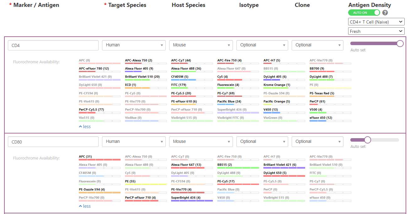 Do you have another feature you would like to see added to FluoroFinder? Contact us to offer suggestions.


