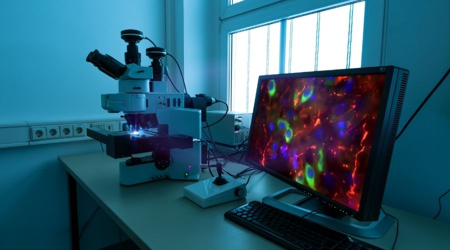
For over 100 years, researchers have used fluorescence microscopy to study biological samples like plated cells or tissue sections. Such experiments typically involve using fluorophore-labeled antibodies to recognize and bind targets of interest before imaging with a fluorescence microscope to produce a magnified view of the specimen. Although fluorescence microscopy is an extremely powerful technology, it is essential to remember that every workflow stage is susceptible to error. This article suggests some key factors to consider when designing a fluorescence microscopy experiment and shares tips for ensuring data accuracy.
Consider the entire workflow
While fluorescence microscopy experiments differ depending on the nature of the sample material and the number and types of biomolecule being studied, they broadly follow a similar workflow. This begins with establishing the biological question to be answered and is followed by identifying suitable sample material and assay conditions. Key considerations here include determining how many samples are required for statistical relevance, defining the region of interest (ROI), and optimizing the sample processing methodology, which spans everything from sample collection, fixation, permeabilization, and blocking to immunostaining and signal amplification. Image acquisition, processing, and analysis must also be considered, and it is important to decide how the data will be presented. To avoid inaccurate reporting, measures should be implemented throughout the entire workflow to safeguard the quality and consistency of results.
Take steps to prevent bias
Operator bias is a well-documented source of experimental variability. For fluorescence microscopy, it can especially be problematic during protocol steps involving visual assessment of images, where the accumulated effect of even small differences can lead to skewed data and ambiguous results. Approaches to avoid operator bias include acquiring images from a predetermined number of locations within a well (or scanning the entire well) when working with plated cells and imaging multiple areas rather than just a single ROI when surveying tissue sections. Other proven strategies involve barcoding samples such that their identity remains unknown during image acquisition and increasing the number of operators acquiring the data.
Ensure accuracy and reproducibility
As well as taking steps to avoid operator bias during image acquisition, it is essential to put in place additional measures designed to ensure data quality. For experimental accuracy, understanding the target of interest is key to having confidence that any imaging data is truly representative of the sample being analyzed. Valuable insights into protein abundance and location can be gained from open-access sites such as PAXdb, UniProt, and the Human Protein Atlas, while mining the available literature can reveal tried and trusted methods for detection. For experimental reproducibility, each protocol step should be meticulously documented such that any operative could follow written instructions to produce comparable results. Recording assay conditions (e.g., ambient temperature, batch numbers of individual reagents, and consumables) during every run supports rapid identification of sources of variability.
Perform rigorous validation
The primary goal of validation is to confirm that the chosen methodology and tools accurately detect the intended target. In terms of fluorescence microscopy, this means assessing antibody reagents for specificity and sensitivity, and determining that the experimental protocol and type of imaging system are suitable for monitoring the analyte(s) in question. Factors to consider include ensuring autofluorescence is not misidentified as the target of interest; confirming primary antibodies still recognize the target following conjugation to a fluorescent label; evaluating the impact of environmental conditions on the behavior of fluorescent proteins, and comparing fluorophore intensities in live and fixed samples to rule out any detrimental effects of using fixative agents. Imaging system validation commonly involves collecting images of known samples, identifying systematic (repeatable) errors present in those samples, and performing error correction – for example, by adjusting the sample processing method or image acquisition parameters.
Include suitable controls
Controls are vital for tracking assay performance and benchmarking results. In addition to known positive and negative sample types, fluorescence microscopy controls include knockout samples (e.g., material produced using CRISPR-Cas gene editing) for confirming antibody specificity; unlabeled controls (samples with no added fluorophore) for evaluating background fluorescence and setting acquisition parameters; single-labeled controls (samples labeled with only one fluorophore) and fluorescence-minus-one (FMO) controls (samples labeled with all but one fluorophore) for measuring bleed through; and secondary antibody only controls (samples where the primary antibody is omitted) for establishing whether secondary reagents exhibit non-specific binding. Photobleaching controls comprise a fluorophore at a steady (unexcited) state and are used to track fluorescence intensity over time. For live-cell imaging studies, experiments performed with no fluorescence illumination are vital to ensure sample material is not damaged by the image acquisition process.
Fluorophore selection
Fluorophore selection must consider key fluorophore characteristics, including the cellular localization and abundance of the target. Fluorophore excitation and emission spectra must be compatible with the chosen microscopy platform to enable detection, and spectral overlap should be avoided to mitigate the risk of generating false-positive results. When monitoring the expression of fluorescent proteins, maturation time (the length of time following translation for the protein to become fluorescent) is often a critical factor; this can be as little as 10 minutes for mNeonGreen, yet over 2 hours for mRuby3. It is recommended that bright fluorophores be paired with rare targets or low-density antigens, and vice versa. Small, organic fluorophores rather than large fluorescent proteins should be selected for detecting intracellular targets where possible since the latter may struggle to cross the cell membrane. Where experiments will take place over an extended time, photostable fluorophores are preferred.
Reagent selection for fluorescence microscopy
With so many different fluorophores available, designing a fluorescence microscopy experiment can seem daunting. However, FluoroFinder is here to help! Our Spectra Viewer lets you quickly compare over 1000 fluorophores from all suppliers in one intuitive platform, many of which have been published for fluorescence microscopy-based research, helping you optimize your multiplexed experiments with the very latest fluorophore and antibody offerings across >100 suppliers. And our technical support team is always available to offer advice and support, whatever the aim of your project.


