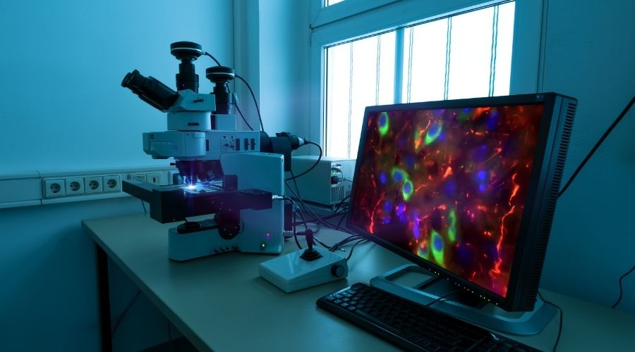
For over 100 years, researchers have used fluorescence microscopy to study biological samples like plated cells or tissue sections. Such experiments typically involve using fluorophore-labeled antibodies to recognize and bind targets of interest before imaging with a...
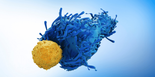
Rare cell types provide novel insights into mechanisms of health and disease Many techniques used for cell-based research rely on bulk analysis, with data being generated from a complex heterogeneous population. The main drawback of these methods is that they can lead...
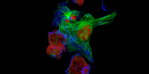
Conventional fluorescence microscopy uses fluorophore-labeled antibodies to visualize cellular structures, providing insights into cellular physiological and pathological states. However, the wave nature of light imposes a resolution limit of approximately 200 nm on...
In recent years, technologies for single-cell analysis have evolved rapidly, revealing huge differences between cells once categorized as being the same type. This variability is most apparent within the cells of the immune system, which must undergo multiple dynamic...
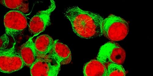
The “optimized multicolor immunofluorescence panel” OMIP publication format was launched 11 years ago as a collaborative platform to establish criteria for experimental design, data collection, and analysis [1]. An OMIP is, by definition, a peer-reviewed publication...
A technique that combines flow cytometry with digital microscopy promises novel insights Imaging flow cytometry (IFC) has seen increased uptake in recent years, largely due to instrumentation and software improvements that have made the technique more accessible. By...