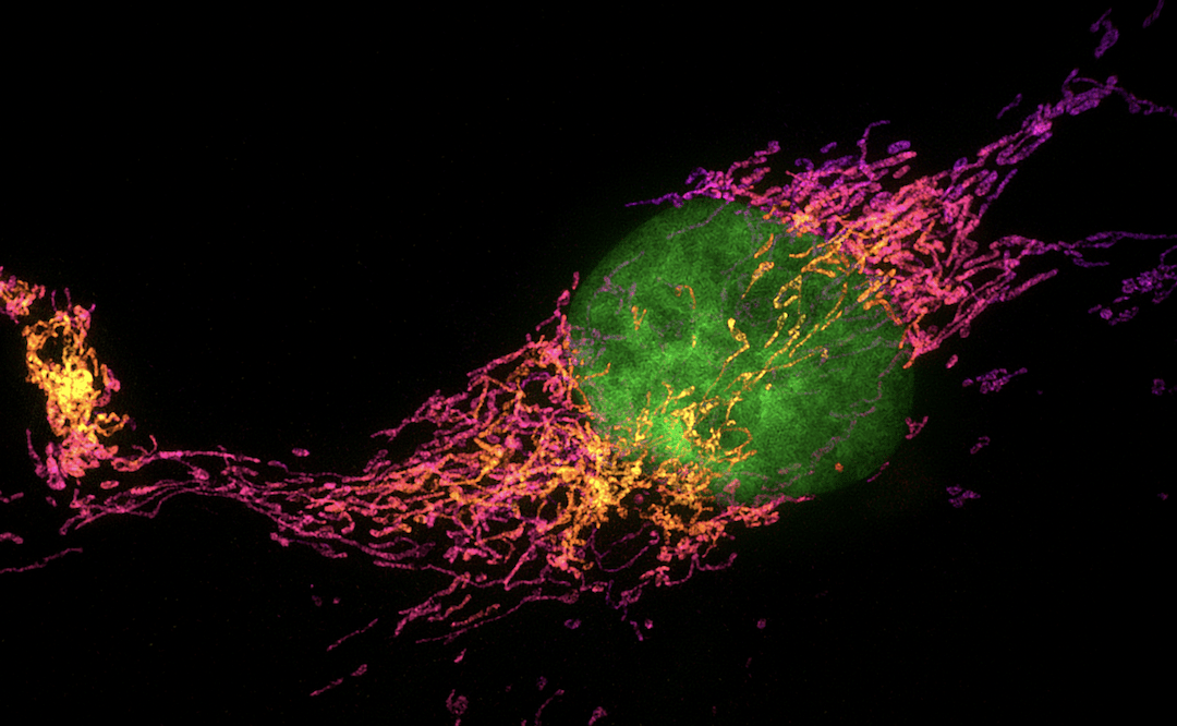
The selection of method and fluorophores for super-resolution microscopy depends on several considerations.
Fluorescence microscopy is one of the most widely used imaging modalities for scientific research owing to its capacity for multiplexed detection and its ability to capture cellular dynamics in real-time. However, light diffraction limits the lateral resolution of conventional fluorescence microscopy to around 200 nm, prohibiting the study of certain cellular features or phenomena. Within the past few decades, various super-resolution microscopy (SRM) techniques have been developed that overcome this constraint. We provide a brief overview of some of the main SRM techniques here, including factors to consider for method selection. In addition, Biotium and Cytoskeleton comment on the importance of collaboration to develop new fluorophore products for SRM applications.
What is Super-Resolution Microscopy?
Super-resolution microscopy refers to a growing array of techniques that break through the optical resolution limit of fluorescence microscopy. This limit is described by Abbe’s law, which states that resolution is equal to λ/(2NA), where λ is the wavelength of light used to illuminate the sample and NA is the numerical aperture of the microscope’s objective. To put this into context, consider that a standard 594 nm laser and an oil-immersion objective with an NA of 1.45 would provide a theoretical resolution of around 205 nm – a value that is considerably greater than the distance between many biomolecules or subcellular structures.
SRM techniques break through the optical resolution limit in one of two main ways. The first of these uses patterned illumination to reduce the size of the point spread function (the point of light and any surrounding blurring emitted by a fluorophore). Examples of these types of methods include:
- Stimulated emission depletion microscopy (STED). First described by Stefan W. Hell and Jan Wichmann in 1994, STED uses two co-aligned beams for sample illumination (1). While one beam excites fluorescence to produce a bright spot, the other provides a donut-shaped pulse of a longer wavelength to bleach the excited fluorophores at the edge of the spot and return them to their ground state.
- Reversible saturable optical linear fluorescence transitions (RESOLFT). RESOLFT generalizes the principles of STED through reversible photoswitching of a fluorescent protein between two conformational states (2). It was demonstrated using the marker protein as FP595 from Anemonia sulcata, which can be switched off and on with blue and yellow light, respectively.
- Structured-illumination microscopy (SIM). Initially performed by Mats Gustafsson in 2000, SIM uses non-uniform illumination (usually in the form of stripes) to excite the sample, with the position and orientation of the light being changed several times during imaging (3). This results in Moiré patterns that can be deconvoluted to produce a super-resolved image.
The second approach takes advantage of single-molecule imaging in solid samples, which was first observed by Moerner and Kador in 1989 (4). Methods within this category include:
- Stochastic optical reconstruction microscopy (STORM). The original STORM methodology, reported in 2006, involved switching a Cy3–Cy5 dye pair between a stable dark state and a fluorescent state using light of different wavelengths (5). By repeating the process for multiple cycles, each switching on a different subset of fluorophores, it was possible to construct a super-resolved image. The technique has since evolved into direct STORM (dSTORM), which uses specialized buffers to drive standard fluorescent dyes into long-lived dark states (6).
- Photo-activated localization microscopy (PALM). First demonstrated by Betzig et al. in 2006, PALM has a similar methodology to STORM, but uses genetically encoded fluorescent proteins instead of synthetic dyes (7).
- DNA-based point accumulation for imaging in nanoscale topography (DNA-PAINT). DNA-PAINT is based on transient binding events rather than stochastic photoswitching. Specifically, oligonucleotide-labeled antibodies are used to bind targets of interest, then the transient binding of fluorescently labeled oligonucleotides with a complementary sequence is measured (8). By eliminating the need for photoswitchable fluorophores or specialized buffers, DNA-PAINT offers more opportunities for multicolor imaging.
It is worth noting that the 2014 Nobel Prize in Chemistry was awarded jointly to Eric Betzig, Stefan W. Hell and William E. Moerner for the development of super-resolved fluorescence microscopy.
Factors to Consider for Method Selection
Depending on the SRM technique that is chosen, researchers may have to compromise on the number of targets detected, the temporal resolution, or the impact of phototoxicity. Key factors to consider are provided in the form of a decision tree in a 2021 Journal of Biological Chemistry publication, which includes the factors listed below (9). In addition, a 2022 Molecular Cell review compares the commercial availability, equipment costs, and relative difficulty of different SRM techniques, which should also be investigated (10).
- The biological information required – structure localization, live-cell dynamics, or molecular interactions
- The type of sample being imaged
- The size of the object of interest
- Whether multicolor detection is necessary
- If single molecule data is needed
Fluorophores for SRM
Each SRM technique requires fluorophores with different properties, meaning that finding the best dye is something that researchers must determine empirically. However, a growing number of publications describes the use of well-known dyes for SRM, and manufacturers can often recommend products for specific methods – including dyes that have been developed through collaboration with SRM experts.
“Of the different techniques described above, Biotium has concentrated specifically on developing dyes and products for dSTORM,” reports Wai-Yee Leung, Ph.D., VP Research and Development. “Our red-excited CF® Dyes, such as CF®647, CF®660C, and CF®680, are extremely bright and have unique photoswitching properties that make them ideal for dSTORM imaging. We have also developed two green-excited CF® Dyes in collaboration with UC Berkeley to advance the potential for dSTORM multiplexing. When compared with other green-excited dSTORM dyes (Alexa Fluor® 532, Cy3B, Atto™ 565, and Alexa Fluor® 568), CF®583R and CF®597R exhibited superior photoswitching under the same excitation power that allowed for high-quality two-color imaging of the challenging structure of actin cytoskeletons.” Full details of this research can be found in a recent Angewandte Chemie publication (11).
Other Biotium products for STORM include Mix-n-Stain™ STORM CF® Dye Antibody Labeling Kits, CF® Dye Single Label Conjugates for STORM, and MemBrite® Fix Cell Surface Staining Kits, as well as ExoBrite™ STORM CTB EV Staining Kits for dSTORM imaging of extracellular vesicles and exosomes. Biotium has also collated a list of publications citing the use of CF® Dyes for various SRM techniques, including STED, STORM, and DNA-PAINT.
Henrick Horita, Ph.D., Marketing Manager at Cytoskeleton, highlights the development of Spirochrome probes, which were tested with STED applications by Hell et al (12). “Spirochrome probes were developed to address the need for high-resolution imaging of the cytoskeleton in living cells,” he says. “They are based on switchable silicon-rhodamine (SiR) derivatives that display an increase in far-red fluorescence intensity upon binding to microtubules or F-actin. Testing included live-cell imaging of human primary dermal fibroblasts, which are notoriously difficult to transfect and therefore challenging to image with genetically encoded probes, and actin staining of intact erythrocytes in whole blood, where the far-red emission of Spirochrome probes is important for minimizing the impact of sample autofluorescence.” Spirochrome probes are also available for imaging lysosomes and chromosomal DNA, and have evolved into next-generation SPY™ Probes that improve on the original technology.
Supporting SRM Research
Selecting the right fluorophores for SRM involves thinking differently compared to selecting fluorophores for conventional fluorescence microscopy – it pays to investigate the range of options available during experiment design. Fortunately, FluoroFinder’s Microscopy Spectra Viewer enables you to quickly compare over 1,300 fluorophores, including many validated for SRM, from all suppliers within one intuitive platform.
References
- https://pubmed.ncbi.nlm.nih.gov/19844443/
- https://www.pnas.org/doi/full/10.1073/pnas.0506010102
- https://pubmed.ncbi.nlm.nih.gov/10810003/
- https://pubmed.ncbi.nlm.nih.gov/10040013/
- https://pubmed.ncbi.nlm.nih.gov/16896339/
- https://pubmed.ncbi.nlm.nih.gov/18646237/
- https://pubmed.ncbi.nlm.nih.gov/16902090/
- https://pubmed.ncbi.nlm.nih.gov/24487583/
- https://pubmed.ncbi.nlm.nih.gov/34015334/
- https://pubmed.ncbi.nlm.nih.gov/35063099/
- https://onlinelibrary.wiley.com/doi/10.1002/anie.202113612
- https://pubmed.ncbi.nlm.nih.gov/24859753/


