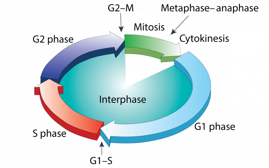
Aberrant cell proliferation and cell death underlie a multitude of disease states Normal tissue homeostasis depends on a critical balance between cell proliferation and cell death. The cell cycle regulates the former, while the latter occurs via controlled...
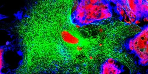
Confocal microscopy is a powerful imaging technique used to study biological specimens. It offers several advantages over conventional widefield microscopy, including the capacity to control depth-of-field and collect serial sections from thick samples. Fluorophores...
Best practices for antibody and fluorophore use safeguard assay performance A defining feature of flow cytometry is its capacity to analyze single cells. This has led to its application across the entire drug development continuum, with recent advances in the field of...
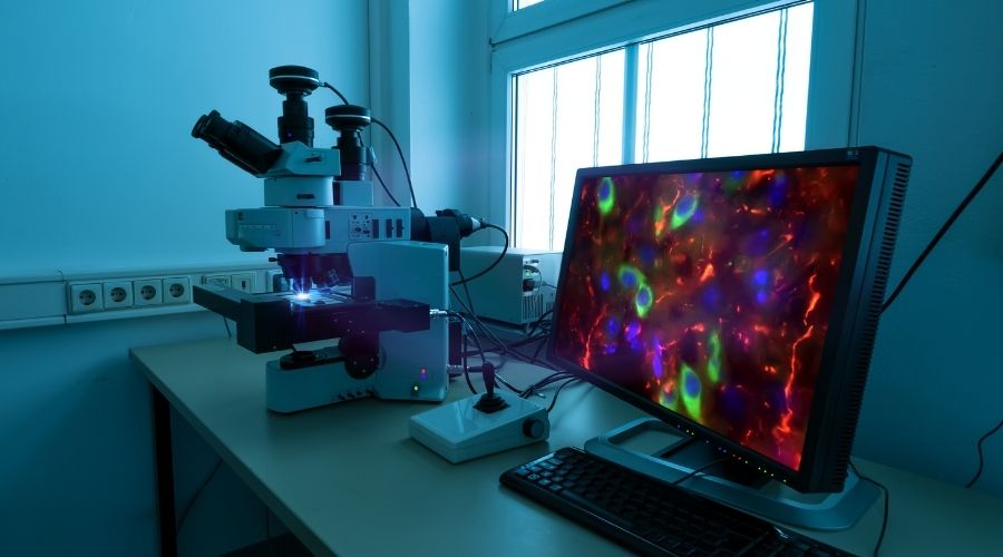
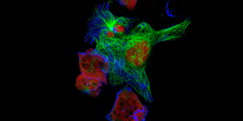
For over 100 years, researchers have used fluorescence microscopy to study biological samples like plated cells or tissue sections. Such experiments typically involve using fluorophore-labeled antibodies to recognize and bind targets of interest before imaging with a...
Rare cell types provide novel insights into mechanisms of health and disease Many techniques used for cell-based research rely on bulk analysis, with data being generated from a complex heterogeneous population. The main drawback of these methods is that they can lead...
Conventional fluorescence microscopy uses fluorophore-labeled antibodies to visualize cellular structures, providing insights into cellular physiological and pathological states. However, the wave nature of light imposes a resolution limit of approximately 200 nm on...