Flow cytometry continues to evolve with advances in instrumentation, reagents, and software The first fluorescence-based flow cytometer was developed in 1968 and was commercialized the following year. Since then, the field of flow cytometry has matured rapidly, and...
Studying calcium flux leads to insights into normal physiology and disease Calcium ions (Ca2+) are involved in numerous intracellular signaling cascades and in a broad range of physiological processes. These include neurotransmission, muscle contraction, and hormone...
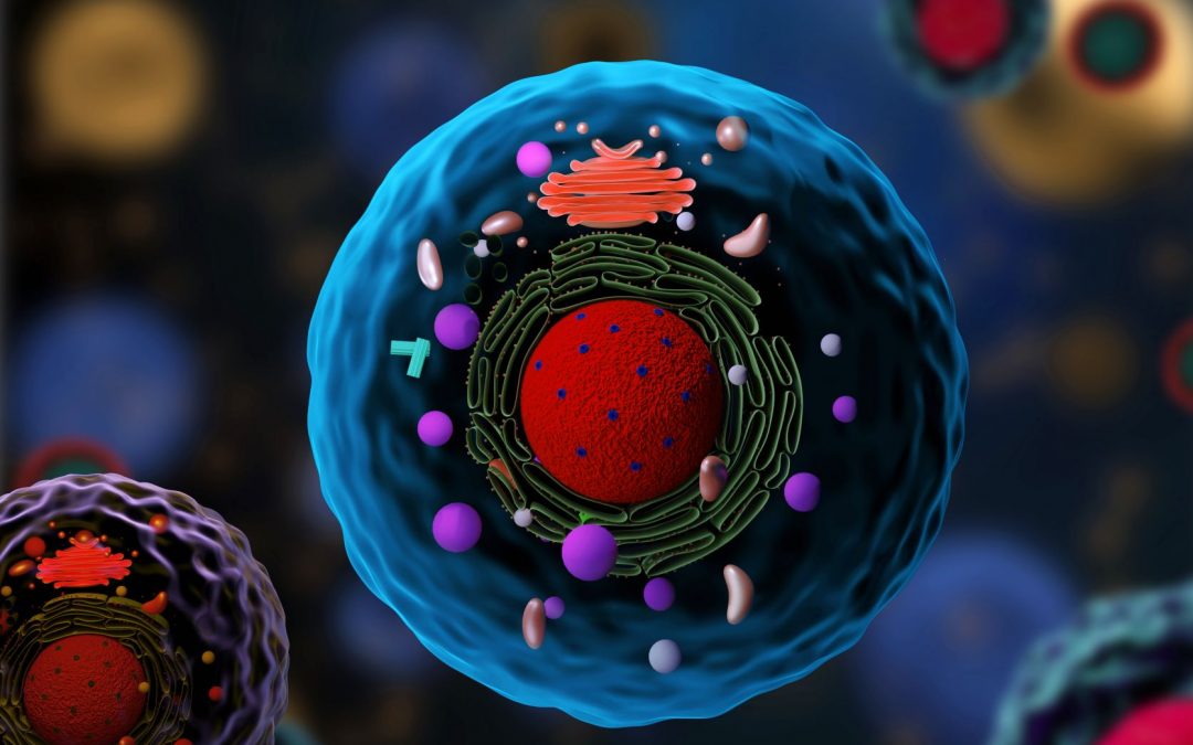
Marker selection and panel design underpin the accuracy of cellular identification Being able to monitor the presence and relative abundance of different cell types in sample material is essential to understand normal biology and investigate the pathogenesis of...
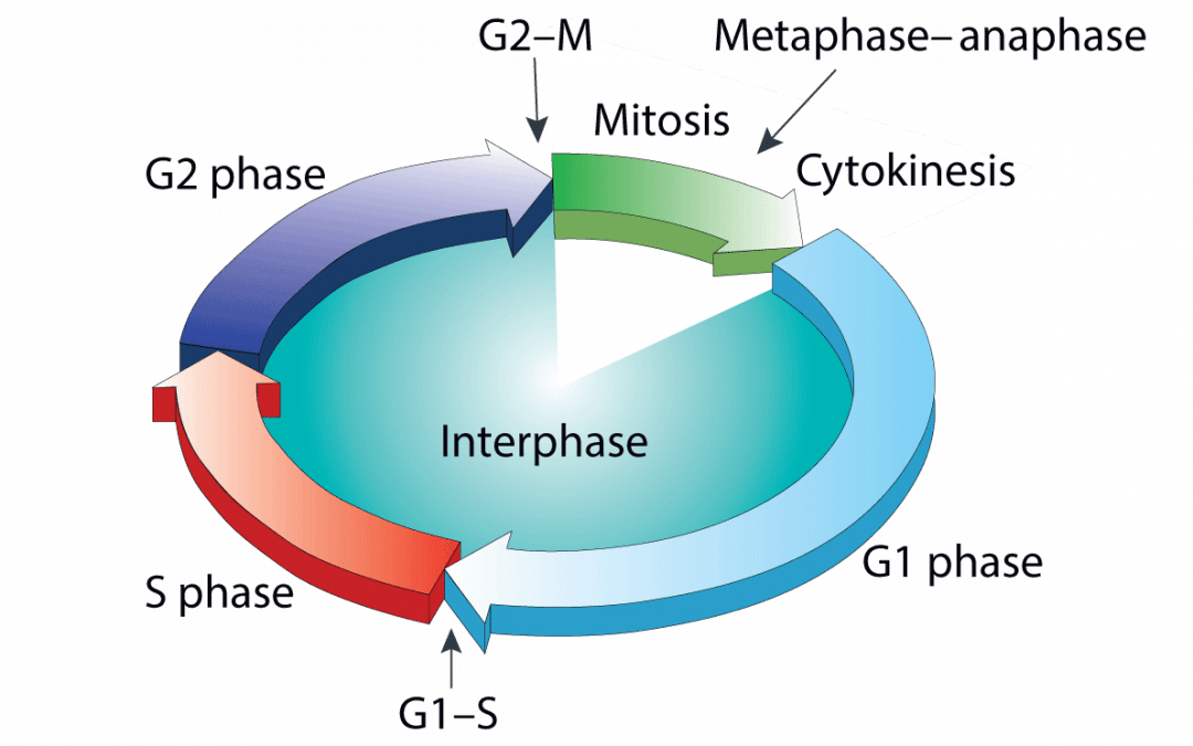
Aberrant cell proliferation and cell death underlie a multitude of disease states Normal tissue homeostasis depends on a critical balance between cell proliferation and cell death. The cell cycle regulates the former, while the latter occurs via controlled...
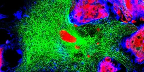
Confocal microscopy is a powerful imaging technique used to study biological specimens. It offers several advantages over conventional widefield microscopy, including the capacity to control depth-of-field and collect serial sections from thick samples. Fluorophores...
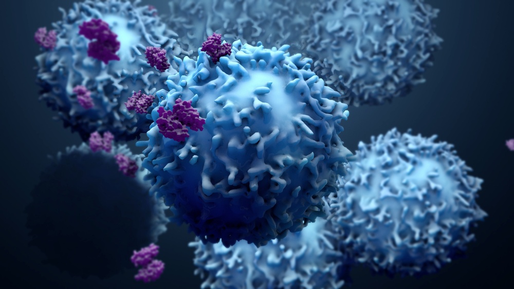
Best practices for antibody and fluorophore use safeguard assay performance A defining feature of flow cytometry is its capacity to analyze single cells. This has led to its application across the entire drug development continuum, with recent advances in the field of...