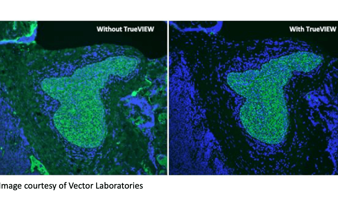
Reducing autofluorescence is critical in fluorescence-based research Techniques such as fluorescence microscopy, flow cytometry, and western blotting often rely on the use of fluorophore-labeled antibodies. The main reason for this is that fluorescence-based...
Fluorescent cellular analytical technologies allow us to “see” beyond what was historically possible with histological stains or morphological scatter profiles. In the early days, microscopy employed excitation sources like arc lamps, isolating specific wavelengths of...
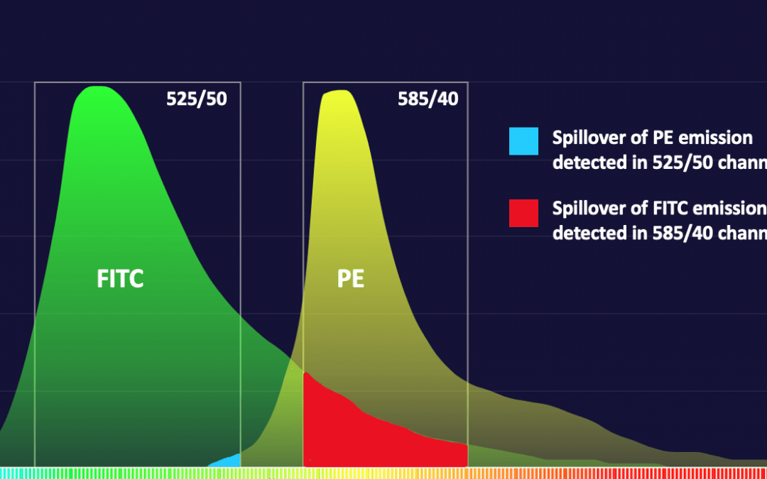
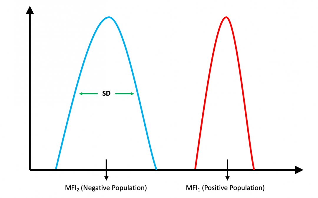
Choosing the right fluorophores is critical for reliable results Fluorescent detection offers significant advantages, including multiplexing capability, superior sensitivity, and a broader dynamic linear range compared to other detection methods. However, the...
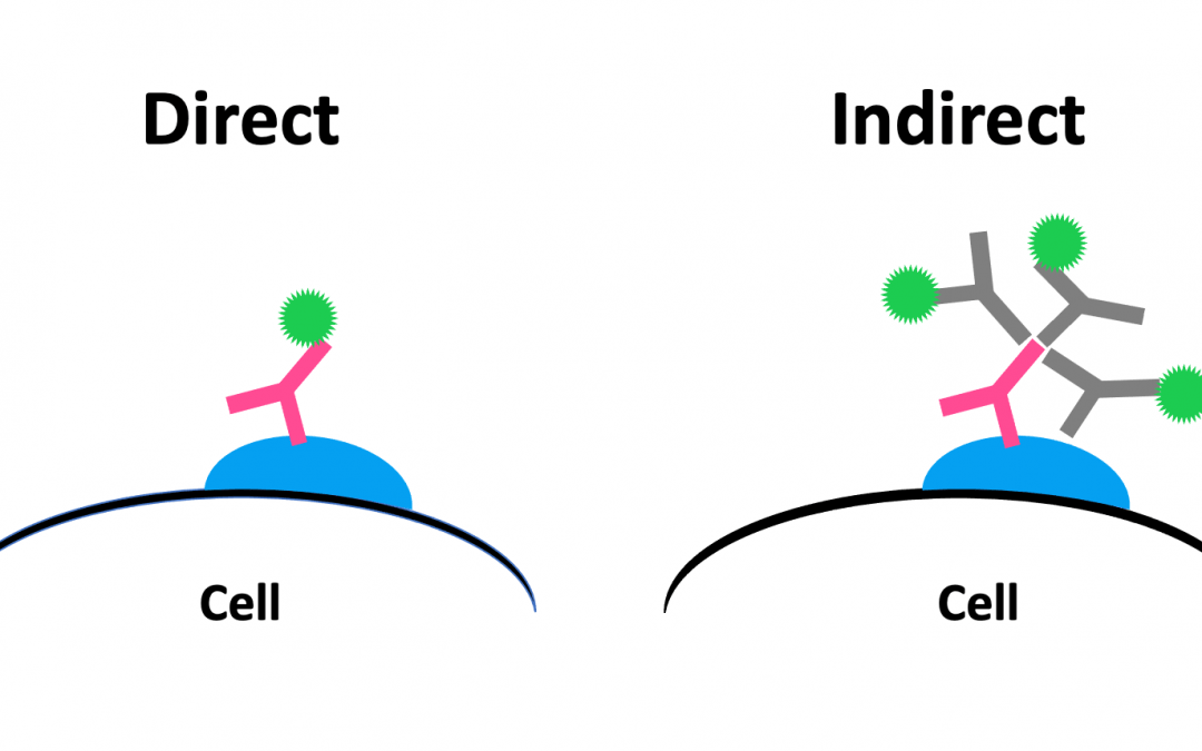
Understanding the advantages and disadvantages of different immunodetection strategies is important to achieve accurate results. Microscopy-based techniques such as immunocytochemistry (ICC) and immunohistochemistry (IHC) often use fluorophore or enzyme-labeled...
When designing a flow cytometry experiment, it is important to account for the relative brightness of each fluorescent label on your specific instrument. Ideally, brighter fluorophores should be assigned to weakly expressed markers, while dimmer fluorophores should...
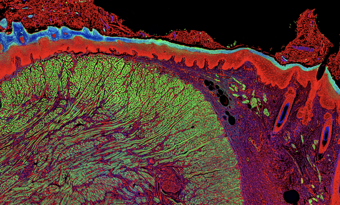
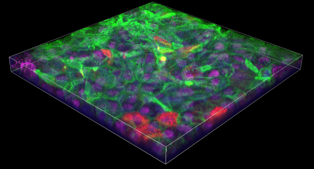
Advanced microscopy techniques enable deeper imaging Advanced microscopy platforms are becoming more widespread for the depth of information they provide. Among these newer modalities, confocal microscopy has risen in popularity for imaging thick...