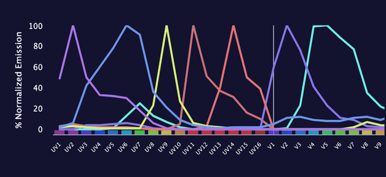
Spectral cytometry offers increased flexibility for fluorophore selection but researchers should still apply best practices for panel design. It has been almost 20 years since spectral cytometry was first described by the Robinson group at Purdue University...
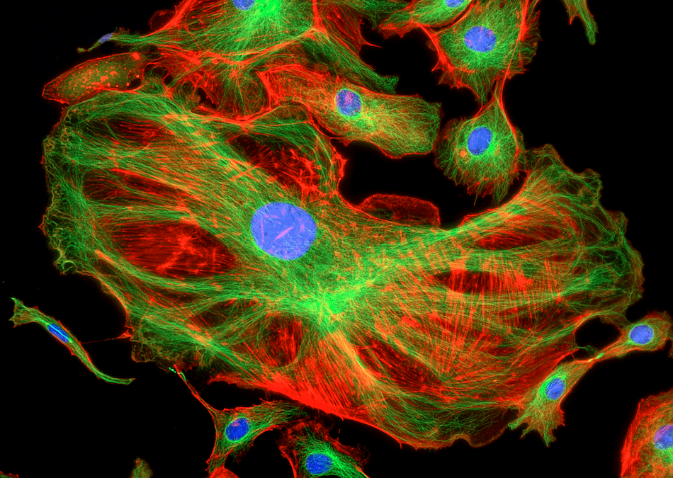
Protocol optimization is essential for clear, reproducible results Fluorescence microscopy uses fluorophore-labeled antibodies and other fluorescent reagents to visualize targets of interest in cells and tissues. Experiments can be as simple as detecting one...
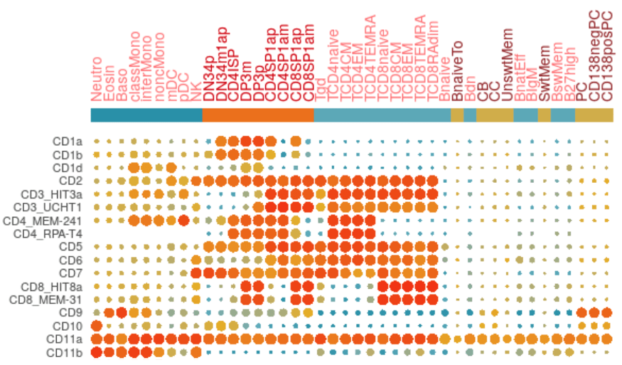
Understanding the biological density of proteins or antigens expressed by each individual cell is an imperative component of all cell-based analytical methods. The comparative change in protein expression, also called antigen density, is indicative of the...
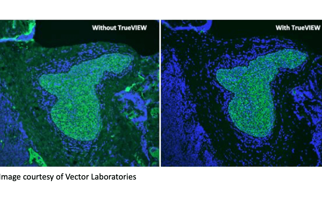
Reducing autofluorescence is critical in fluorescence-based research Techniques such as fluorescence microscopy, flow cytometry, and western blotting often rely on the use of fluorophore-labeled antibodies. The main reason for this is that fluorescence-based...
Fluorescent cellular analytical technologies allow us to “see” beyond what was historically possible with histological stains or morphological scatter profiles. In the early days, microscopy employed excitation sources like arc lamps, isolating specific wavelengths of...
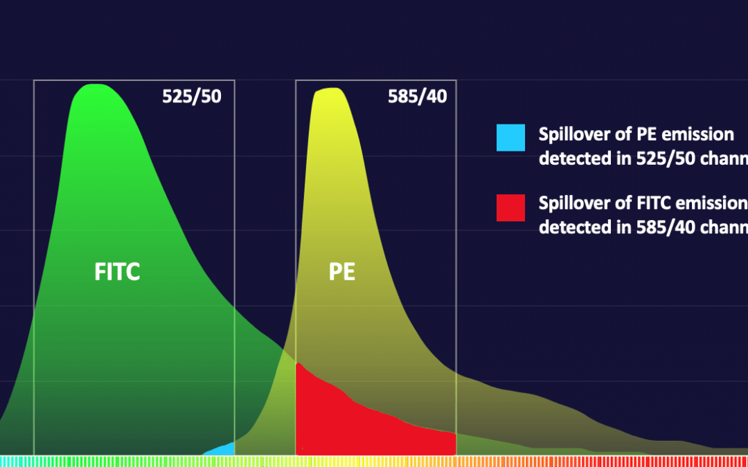
Choosing the right fluorophores is critical for reliable results Fluorescent detection offers significant advantages, including multiplexing capability, superior sensitivity, and a broader dynamic linear range compared to other detection methods. However, the...