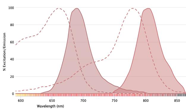
Near-infrared fluorophores offer several advantages for imaging applications. A major advantage of fluorescent detection is that it enables multiple antibodies to be combined in the same experiment. Using techniques such as immunocytochemistry (ICC) and...
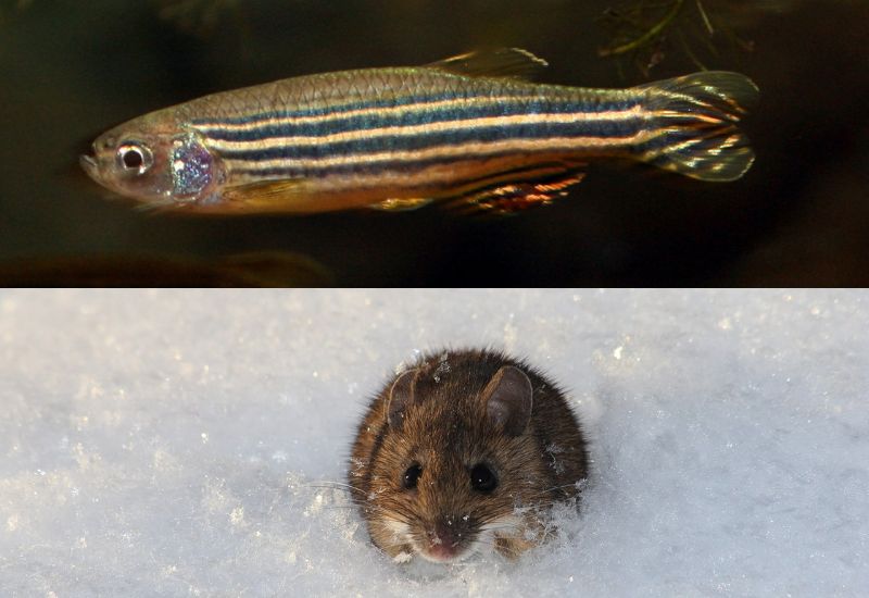
Fluorescence microscopy is seeing increased utility for imaging organisms While fluorescence microscopy has long been employed for imaging plated mammalian cells, its use for visualizing organisms is flourishing. In recent years, winning images in Nikon’s annual Small...
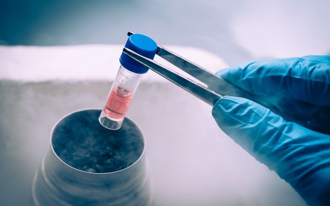
High viabilities, well-preserved antigens, and minimal clumping underpin flow cytometry success Unlike bulk population analysis techniques such as ELISA and Western blot, flow cytometry provides information about individual cells. Although microscopy does this as...
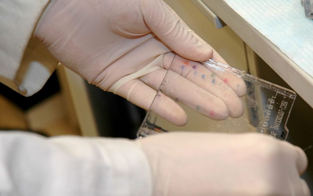
What are OMIPs? Optimized multicolor immunofluorescence panels, commonly known as OMIPs, are highly validated sets of antibodies and reagents that are published, peer-reviewed, and compiled to serve as a resource to the scientific community. OMIP publications often...
New approaches to Western blotting techniques overcome performance limitations For over 40 years, researchers have used Western blot for protein identification. Not only does Western Blot provide confirmation that a target of interest is present in a sample, this...
Expansion Microscopy physically expands biological specimens enabling higher resolution imaging First described in 2015, expansion microscopy is an imaging technique that improves on the resolution of conventional light microscopy by physically expanding the...