Written by Sarah Locknar
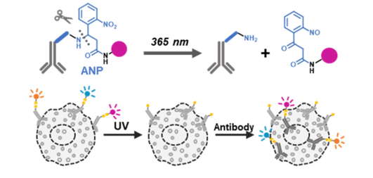
Cover: The PACIFIC method of iterative multiplexing uses photocleavage between labeling cycles. Used without modification under Creative Commons Attribution 4.0 International License from [1], graphical abstract.
Iterative or Cyclical Multiplexed Immunofluorescence Methods
The easiest way to perform high-plex immunofluorescence imaging without upgrading hardware (to include additional lasers, spectral detection or multimodal capabilities) is to perform sequential, iterative or cyclical labeling protocols. The widespread deployment of integrated imaging systems with microfluidic chambers has enabled these protocols to be performed automatically without intervention, or manually for a cheaper option.
The general workflow for these protocols includes:
- Tissue preparation (usually FFPE or frozen tissue sections) adhered to a glass substrate followed by deparaffinization and rehydration and antigen retrieval (if required)
- Blocking
- Antibody staining
- Nuclear counterstaining
- Imaging
- Signal removal
- Postprocessing image registration – using nuclear staining as fiducials
- Postprocessing background subtraction
- Postprocessing quantification of expression levels
- Postprocessing image analysis, including cell segmentation, colocalization assessment, etc.
Steps 3-10 are repeated to achieve data sets with 50 or more labels. Most of the differences in techniques involve steps 3 and 6, as described in the following sections. Methods can be loosely grouped into those that use bleaching between labeling rounds and those that use dissociation or stripping.
Iterative multiplexing by bleaching or inactivation
CmIF CycIF, [2] tissue CycIF (t-CycIF), [3], MxIF [4] and IBEX (iterative bleaching extends multiplexity) [5] use fluorescently-labeled primary antibodies that remain in place. After imaging, the fluorophores are bleached using various methods. CycIF and t-CycIF use base-catalyzed photo-oxidation with sodium hydroxide and peroxide [2] [3]. CmIF [6] uses photobleaching and MxIF. [4] utilizes base-catalyzed oxidation with heating. IBEX inactivates the fluorophore by reduction with lithium borohydride [5].
Not all fluorophores are easily bleached with each method, so care should be used when selecting dyes. Bleaching efficiency can be assessed by imaging the tissue at different timepoints during the bleaching protocol. Using a flow-chamber or successive washes with bleaching solution can also improve bleaching rates. [5] It is important to thoroughly rinse out the bleaching solution so as not to affect fluorescence signal in the next labeling cycle. Successive rounds of bleaching can have the benefit of reducing autofluorescence and increasing the signal-to-noise over time. [3]
Iterative multiplexing by stripping or dissociation
In these methods, the antibodies or fluorescent labels are denatured and/or dissociated from the binding sites and washed away between staining steps.
The SeqIFTM protocol [7] strips primary and secondary complexes off the tissue using an acidic detergent solution. Lunaphore (now Biotechne) supplies this elution buffer, designed to be used in their CometTM system.
The 4i technique (iterative indirect immunofluorescence imaging) [8] uses off-the-shelf primary and secondary antibodies with a radical scavenging imaging buffer to reduce photo-induced crosslinking between antibodies and epitopes during the imaging process. Imaging is followed by acidic guanidinium hydrochloride and urea treatment to denature and release the antibodies.
Quanterix’s (formerly Akoya) PhenoCycler®-Fusion (previously known as CODEX®) is designed to work with oligonucleotide barcode-conjugated antibodies. Oligo-conjugated reagents are available from Quanterix, or the conjugation can be done independently [9]. At the beginning of the experiment, antibodies against a large number of different antigens are bound to the sample (up to 100 have been demonstrated). During a staining cycle, fluorescently tagged oligo “reporters” hybridize to the complementary subset of bound antibodies. After imaging, the reporters are released with a high salt solution. [9] A different subset of antibodies are labeled in each subsequent staining cycle.
SeqStain uses fluorescent DNA-conjugated primary or secondary antibodies for labeling and imaging, followed by an enzymatic nuclease cleavage step to remove fluorophores between cycles. [10] This protocol allows for some amount of amplification and flexibility. Typically, 2-5 docking oligonucleotides are conjugated to each antibody. The docking oligonucleotide can contain a number of docking sequences (enabling binding of multiple fluorescent DNA complexes) and each fluorescent DNA complex can contain more than one fluorophore. [10]
PACIFIC (photoactive immunofluorescence with iterative cleavage, cover figure) [1] uses a photocleavable linker (amino-3-(2-nitrophenyl)propionic acid, ANP) to attach fluorophores to antibodies. After irradiation at 365 nm for 1 h, the fluorescent fragment is released. To help with solubility of the fluorescent fragment, the linker includes a poly-ethylene glycol moiety. [1]
Setting up your iterative or sequential labeling experimente
Preparing the sample
Thin sections (<10 micron) of FFPE tissues followed by antigen retrieval methods like heat-induced epitope retrieval (HIER), or cryo-sections are commonly used. Thicker sections require much longer processing times and 3-D imaging techniques like confocal or light-sheet microscopy. Tissues with high autofluorescence can be photobleached or quenched with chemical reagents before labeling. To learn more, see our article “Increasing signal to noise ratio in fluorescence microscopy and blocking methods”.
Immobilizing the sample
To facilitate the registration of images from each cycle, it’s crucial to immobilize the sample onto the substrate. Slides with a positively charged coating, such as SuperFrost or poly-l-lysine, are often used for standard histological analysis using H&E or single-cycle immunofluorescence. FFPE slices are adhered with heating at 65o C. This approach is not recommended for protocols that require cyclic heating, as slices have been seen to lift off. [11] In contrast, UV-induced cross-linking robustly attaches FFPE sections onto glass and preserves the structure through many rounds of heat treatment. [11] Silylated slides, available from a number of manufacturers including Sigma-Aldrich and Thomas Scientific, covalently bond with tissue slices and exhibit low autofluorescence.
For cryosections, gelatin subbed slides are often used. These are available from suppliers like Fisher Scientific. These slides use a crosslinker (chromium potassium sulfate) to bond the gelatin and tissue to the glass surface. One drawback of gelatin is possible autofluorescence. Another method uses Cryotape to hold cryosections followed by adhering the back of the tape to glass substrates with UV-cure optical epoxy. [12]
Per cycle planning
Per-cycle planning depends on your hardware (number of excitation wavelengths, number of emission filters or spectral unmixing capabilities) and how many signals can be successfully resolved in one labeling cycle (usually 3-6). Knowing this limit will help in designing the rest of the experiment. In many ways, the experimental design is similar to a series of single-cycle multiplexed experiments, keeping in mind that the sample may change over time. For instance, background signal may change, and in some cases, antigens may be destroyed between cycles.
Typical immunofluorescence controls are required for good results including single-antibody testing and titration, isotype controls, positive and negative controls. It is also important to verify that the signal is destroyed before moving on to the next cycle. This can be checked by imaging between cycles to see if any fluorescence remains. Antibody elution protocols like SeqIFTM and 4i, require controls using no primary antibody between cycles to confirm complete removal.
Dye Choice
Dyes should be chosen strategically so that they are compatible with your inactivation chemistry and your hardware. The same fluorophores can be used repeatedly in different cycles to label different antigens.
The brightest, most photostable dyes should be used for the least abundant targets. Resources such as PAXdb and The Human Protein Atlas can help researchers determine concentrations of various antigens in their samples.
Mitigating epitope loss
Loss of epitopes can occur with many of the chemical bleaching methods, especially those that involve heating. Some of the antibody stripping methods are also quite harsh – high salt and detergents can denature and solubilize some proteins (and epitopes) along with the antibodies. The enzyme-based methods are the most likely to preserve sensitive epitopes. Controls that compare results across multiple staining cycles and with single-cycle immunofluorescence can be used to assess epitope retention.
Preparing the sample
Typically, nuclear stains are used to align and overlay images from different staining cycles. Usually, nuclei are restained with each cycle to maintain a robust signal.
Summary
Iterative and sequential multiplexing have greatly expanded the microscopist’s toolbox. Although tricky to set up, with the right controls, ultra high-plex experiments are becoming routine, even for clinical applications. One benefit of these methods is that the same slices can be treated with H&E stain for traditional pathology assessments after the fluorescence data is acquired. This allows researchers and clinicians to understand human tissues like never before.
References
- Ji, 2023. DOI: 10.1021/acsbiomedchemau.3c00018
- Lin, 2015, DOI: 10.1038/ncomms9390
- Lin, 2018, DOI: 10.7554/eLife.31657
- Gerdes, 2013, DOI: 10.1073/pnas.1300136110
- Radtke, 2022, DOI: 10.1038/s41596-021-00644-9
- Eng, 2020. DOI: 10.1007/978-1-4939-9773-2_24
- Rivest, 2023, DOI: 10.1038/s41598-023-43435-w
- Kramer, 2023, DOI: 10.21769/BioProtoc.4712
- Black, 2021, DOI: 10.1038/s41596-021-00556-8
- Rajagopalan, 2021, DOI: 10.1016/j.crmeth.2021.100006
- Zhang, 2024, DOI: 10.1089/ten.tec.2024.0223
- Dyment, 2016, DOI: 10.3791/54468 (2016)