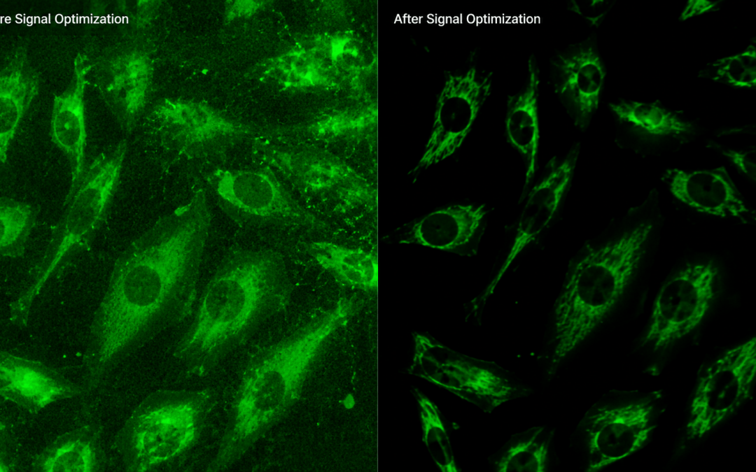
Written by Sarah Locknar
Blocking and signal-to-noise enhancement in immunofluorescence imaging
Advanced fluorescence microscopy techniques like TIRF, STORM and PALM, depend on high signal-to-noise ratios (SNR) to detect small signals down to single-molecules. In this article, we discuss methods to control sources of high background signal arising from autofluorescence, non-specific antibody binding and hardware configurations, to detect the weakest signals.
Autofluorescence background signal
Autofluorescence is the natural fluorescence present in a sample. It is highly dependent on the type of tissue and its preparation method. Live and frozen tissues can contain a number of fluorescent molecules including FAD, NADH, lipofuscin, chlorophyll, tryptophan, phenylalanine, collagen, etc. Formalin-fixed tissues often have autofluorescence spectra that arises from some of the same molecules present in live tissues along with artifacts from protein crosslinking. There are several approaches to reducing autofluorescence, including direct methods using bleaching or chemicals, time-resolved imaging and post-processing subtraction techniques.
Directly reducing autofluorescence
A number of methods for reducing autofluorescence have been described in the literature including broad-spectrum bleaching prior to immunofluorescence labelling (Sun, 2017), ammonia/ethanol treatment, Copper (II) sulfate, Sudan Black B, Trypan Blue and Biotium’s TrueBlack®. (Yang, 2017) (Wu, 2021)
Ammonia/ethanol solutions and Copper (II) sulfate reduce autofluorescence through chemical means. Ammonia is a known denaturing agent which can break up fluorophores and alter the protonation state of protein sidechains, while ethanol can help dissolve chromophores. Copper (II) sulfate solutions likely work by coordination of the Cu2+ to fluorescent moieties leading to chemical quenching via energy transfer. (Hotzer, 2011) Both Trypan Blue (Figure 1a) and Copper (II) sulfate also help reduce autofluorescence by absorbing excitation and emission light in much of the visible spectrum (Figure 1b). Vector Labs offers TrueVIEW autofluorescence quencher which reduces autofluorescence from fixation, collagen, elastin and red blood cells.
Sudan Black B (SBB) and TrueBlack® are hydrophobic molecules (Figure 1a) that preferentially associate with lipid rich autofluorescent vesicles (lipofuscin). They must be dissolved in ethanol or other aprotic solvents like DMSO before use. Given their wide visible absorption spectra (Figure 1b), they strongly absorb both excitation and emission light, reducing lipofuscin and other autofluorescence. SBB’s absorption spectrum is also sensitive to solvent polarity, shifting in wavelength depending on its location within the cell, enabling it to absorb over a wide range (Figure 1b). SBB exhibits significant emission in the near-infrared (above about 650 nm) which led to the development of TrueBlack®. TrueBlack® Plus was developed to increase water solubility and further reduce NIR fluorescence signals. Water solubility also reduces sample shrinkage and enables longer incubation times.
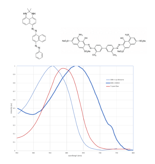
Figure 1. (A) Chemical structures of Sudan Black B (left) and Trypan Blue (right). (B) absorption spectra of SBB (blue) and Tryptan Blue (red). The absorption of SBB shifts to the blue in hydrophobic solvents like cyclohexane.
Removing autofluorescence with FLIM
Fluorescence lifetime imaging (FLIM) uses time to differentiate molecules with similar or overlapping excitation and emission spectra. Each fluorophore exhibits a characteristic decay curve related to how long it stays in the excited state before emitting a photon, which is unique to each fluorophore and its environment. FLIM uses a fast excitation pulse while detecting the fluorescence emission decay between pulses. Typically, emission signal is time-gated into several bins to calculate and display fluorophores with different fluorescence lifetimes, including autofluorescence signal, which can then be removed in post-processing.
Removing autofluorescence with post-processing
Spectral imaging systems allow users to save spectra of autofluorescence of their unlabeled samples that can be subtracted from the labeled images in post-processing. In automated microscopes with good mechanical systems and motors, autofluorescence of the identical field of view can be imaged before immunofluorescence labelling for subtraction later, or autofluorescence spectra can be saved from an unlabeled region of interest and subtracted from the image as a whole.
Background signal from non-specific binding
Non-specific binding occurs when antibodies bind to unintended targets, raising the overall signal of the sample in an unpredictable way. Some researchers believe non-specific binding is not a significant problem (Buchwalow, 2011) but it is surely dependent on the tissue type and its preparation method with respect to fixation, permeabilization and epitope recovery. Non-specific binding can occur via antibody-protein interactions or by charge-based adsorption between negatively charged dye-labeled antibodies and positively charged cellular components. Depending on the main source of background signal, a combination of blocking agents may be required.
Blocking protein binding
Non-specific antibody-protein interactions can be blocked by treating fixed tissues with relatively high concentrations (10-20%) of non-fat powdered milk (Gajda, 2008), bovine serum albumin (BSA), gelatin or casein prior to incubation with primary antibodies. A more specific approach uses normal serum from the same animal as the labelled secondary antibodies. Cell Signaling Technology offers a goat serum based protein blocker with a permeabilization buffer in their Immunofluorescence Blocking Buffer #12411. Active Motif uses a non-mammalian blocking agent in their MAXblockTM product that does not cross-react with secondary antibodies. Newer approaches use copolymers of N-2-(hydroxypropyl)methacrylamide or poly(oxazoline) as alternatives to BSA. (Subr, 2024) The use of synthetic polymers reduces batch-to-batch variability of serums.
Using a mouse model system with mouse secondary antibodies requires extra blocking because both the primary antibody of interest and native mouse antibodies are labeled with secondary antibodies. Nonspecific protein blocking described above is followed by blocking with commercially available anti-mouse IgG blocking reagents to provide the best results. (Bassiouni, 2020) Vector Labs offers blocking serums from a variety of animals, including mouse on mouse blocking.
Blocking charge-charge interactions
Designed specifically to mask nonspecific charge-based adsorption, ThermoFisher’s Image-iTTM FX signal enhancer can be used before a protein blocker. Biotium TrueBlack® IF background supressor and blocking buffer blocks both charge-based adsorption and non-specific protein binding using non-mammalian blocking agents. It also includes permeabilization detergents to help increase antibody penetration and labeling rates.
Background signal from hardware choices
As with all fluorescence experiments, the properties of the fluorophores must be compatible with the imaging system hardware. FluoroFinder’s Microscopy Spectra Viewer will help you ensure that the light sources, filters and detectors are optimized to minimize spectral bleedthrough or crosstalk between fluorescence channels.
Special considerations should be made when working outside of the visible range. Transmission of objective lenses and detector response curves fall off in UV and NIR wavelengths and sometimes unidentified UV and/or IR blocking filters are installed in the illumination path reducing overall signal.
Signal Amplification
Even after optimizing the factors mentioned above, fluorescence signal can still be small; especially when the antigen concentrations are low. In this case, signal amplification techniques can be employed which increase the number of fluorophores at each labeling site. These techniques include indirect detection using primary and labeled secondary antibodies, labeled-streptavidin biotin (LSAB), and tyramide signal amplification (TSA) as described in this article. A relatively new technique called fluorescent signal amplification via cyclic staining of target molecules (FRACTAL) (Yeon, 2022) uses multiple rounds of fluorescently tagged antibodies to increase the signal (Figure 2). The pairs of fluorescent secondary antibodies can be alternated to reduce the number of required reagents. When labelling more than one antigen using this method, extensive antibody purification by cross-adsorption is required to get good results. (Yeon, 2022)
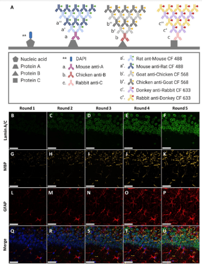
Figure 2. Simultaneous multiplexed IF signal amplification via multiplexed FRACTAL in a mouse brain slice. (A) Schematics of the staining process of the multiplexed FRACTAL with imaging results shown in (B–U). (B)– (U) Multiplexed IF signal amplification imaging of the CA1 region of a mouse brain. (B–F) Signal amplification of lamin A/C. (G–K) Signal amplification of MBP. (L–P) Signal amplification of GFAP. (Q–U) Merge images of (B–P). (Q) Merge image of (B), (G), and (L); Round 1. (R) Merge image of (C), (H), and (M); Round 2. (S) Merge Image of D, I, and N; Round 3. (T) Merge Image of (E), (J), and (O); Round 4. (U) Merge image of (F), (K), and (P); Round 5. Scale bar: 30 μm. Used without modification under Creative Commons Attribution 4.0 International License from (Yeon, 2022) Figure 3.
Improving SNR with image processing
Integrating and averaging during image acquisition can increase SNR because the signal is stationary while non-bound signal tends to be moving or random. Post-processing deconvolution of fluorescence images can also improve SNR by mathematically reassigning photons to a different position in the image based on the known optical properties of the system. Another noise reduction post-processing method termed NEDD (noise estimation, denoising and deblurring) has also recently been described. (Azzari, 2024)
Conclusion
There are many options for improving SNR in immunofluorescence microscopy. Since background signal is highly dependent on tissue type and its preparation methods, a combination of techniques, including autofluorescence removal, blocking of non-specific antibody binding, fluorescence amplification, hardware optimization and post-processing may be needed to provide optimal results.
References
Azzari, V. N. (2024). doi:10.21769/bioprotoc.5072
Bassiouni, S. S. (2020). doi:10.1002/CPMO.84
Buchwalow, S. B. (2011). doi:10.1038/srep00028
Gajda, J. M. (2008). doi:10.2478/V10042-008-0021-8
Hotzer, I. B. (2011). doi:10.1111/j.1742-4658.2011.08434.x
Subr, K. P. (2024). doi:10.3390/polym16060758
Sun, I. C. (2017). doi:10.3791/56188
Wu, F. H. (2021). doi:10.1186/s13071-021-05027-3
Yang, Y. C. (2017). doi:10.12688/wellcomeopenres.12251.1
Yeon, C. S. (2022). doi:10.1038/s41598-022-12808-y