Written by Sarah Locknar
Reagents for Super-resolution microscopy
Super-resolution microscopy (SRM) methods have taken the microscopy world by storm (literally!). These techniques enable imaging an order of magnitude beyond the optical “Raleigh limit”. There are two basic approaches to this technology- (1) single molecule localization microscopy (SMLM) methods which use various techniques (STORM, PALM, DNA-PAINT) to sequentially excite a small subset of fluorophores, building up the image over time, and (2) emission-suppression methods like stimulated emission depletion (STED) that reduce out-of-focus emission in the time domain. Achieving these amazing resolutions requires not only hardware and software, but specific reagents.
One general limitation of SRM is the size of fluorescently tagged antibodies commonly used for labeling. A standard IgG antibody is roughly 10 nm in diameter, which is similar to the resolution limit of these techniques. Image quality can be improved by using VHH antibodies in the 2-4 nm diameter range, available from Biotium (MiniMabTM), Fortis Life Sciences, Thermo Fisher, and Sanofi (Nanobody®).
Single molecule localization microscopy (SMLM)
SMLM methods use dyes that cycle between dark (off) and light (on) states with a low duty cycle (off>>on) so that only a few fluorophores are “on” at a time. [2] Software reconstructs the image after several illumination cycles. The “blinking” can be achieved directly, by adjusting the buffer composition in dSTORM (direct stochastic optical resolution microscopy), by using a reporter and activator pair (STORM), by using activatable fluorogenic probes in PALM (photo activated localization microscopy) or by transient binding with DNA-PAINT techniques.
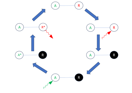
Figure 1. Activator-Reporter based STORM process. Clockwise from top, activator (A) and reporter (R) are close together or linked. A red photon traps the reporter in a dark state (black), absorption of a green photon by the activator excites the activator (A*) and energy is transferred to the reporter resulting in an excited reporter (R*) which emits a photon.
Reagents for STORM and dSTORM imaging
Early STORM methods used activator/reporter dye pairs. [3] Activator fluorophores are chosen to have an emission spectrum that overlaps the reporter absorption spectrum. When an activator fluorophore absorbs a photon within a few nm of a “dark” reporter, (Figure 1) it can transfer its energy to the reporter, turning the reporter on. Activator excitation is performed with low light intensity so as to only activate a small number of dyes at a time. Proximity of activators and reporters can be achieved by directly tethering the dye pairs or binding both to the same antibody or cellular structure. [3] At least two laser wavelengths are required – a long-wavelength laser (Figure 1, red photon) to generate the reporter dark state and a lower wavelength laser to excite the activator (Figure 1, green photon) and initiate reporter emission. This method has the advantage of control of emission cycling and simple spectral multiplexing (using the same activator with spectrally-distinct reporters). [2] [4]
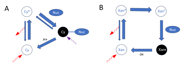
Figure 2. Proposed mechanisms for low-duty-cycle blinking in dSTORM. (A) Cyanine dye in the excited state (*) forms a dark adduct with the nucleophile. The adduct is broken with acid-catalyzed photocleavage from absorption of one UV, or two red photons. (B) Xanthene-based dyes form a dark radical from photoreduction of the triplet state(T). The radical is oxidized to return the fluorescent form.
Evaluating fluorochrome assignments within the context of a specific instrument helps validate panel design before data acquisition. Comparing excitation and emission spectra while assessing spectral overlap, spillover, and fluorochrome compatibility can improve panel performance and reduce the need for iterative redesign.
Reagents for STORM and dSTORM imaging
dSTORM refers to “direct” STORM which doesn’t require energy transfer between dyes. Instead, it utilizes intrinsic low duty cycle blinking [4] which is achieved using readily available cyanine-based dyes (Cy5, Cy7, some of Themo Fisher’s AlexaFluor and Biotium’s CF®-dyes) or xanthene-based fluorophores (rhodamine, Leica’s ATTO, some of Thermo Fisher’s AlexaFluor, Bio-Techne’s Janelia Fluor®, Abberior’s FLUX). [2] In long-chain cyanine dyes, photoinduced electron transfer from a strong nucleophile [5] [6] to the dye in the excited state leads to a long-lived dark dye-nuc adduct (Figure 2A). The dye-nuc adduct undergoes an acid-catalyzed photocleavage to return the ground-state dye [6]. The degree of nucleophilicity, steric hindrance and binding energy determine the dark state lifetime. Stronger nucleophiles bind for a longer time, as do compounds with less steric hindrance. Weak binding energy enables fast on-rates and rapid photoswitching cycles. [5]
For xanthene-based fluorophores (Figure 2B), the nucleophile induces photoreduction of the triplet state to form a dark radical instead of forming a dye-nuc adduct. The radical is reoxidized to reach the ground state. [2] Other xanthene-based fluorophores undergo self-blinking by shifting between “on” zwitterionic and “off” spirocyclized forms. [7] Silicon-substituted rhodamine (SiR) derivatives have higher membrane permeability and longer-wavelength red-near-IR spectra; [8] [9] helpful for multiplexing and live-cell experiments. SiR fluorophores are available from a number of manufacturers including Spirochrome and Lumiprobe.
STORM multiplexing can be achieved with sequential staining via DNA-PAINT techniques, wavelength multiplexing, or frequency multiplexing (using different excitation laser modulation rates). Dye selection should be mindful of the laser wavelengths installed on your system so that all the dyes can be addressed with your hardware. FluoroFinder makes it easy with a comprehensive dye database and interactive spectra viewer to help you choose fluorophore combinations that will work with your installed lasers.
Buffers used in dSTORM experiments are designed to steer the fluorophore into fluorescent or dark states instead of irreversible photobleaching. They tend to have neutral or high pH (~8) to shift the equilibrium towards the dark state (Figure 2a), and contain oxygen scavengers (e.g. ProLong®, Trolox, ascorbic acid, etc.) to ensure that any photo-oxidized fluorophores are reduced back to the ground state instead of bleaching. GLOX, an enzymatic buffer containing glucose, glucose oxidase and catalase (Figure 3), is commonly used in low oxygen imaging, but high glucose concentrations can cause osmotic stress in live cells, and the pH tends to decrease over time. Another approach uses Oxyrase® vesicles (OxyFluor®) which convert dissolved lactate and O2 to pyruvate and water without pH changes. Several other buffer systems have also been described. [10] [11] Care should be taken to reduce hypoxia in live-cell imaging using low-oxygen buffers.
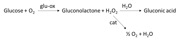
Figure 3. The GLOX buffer uses glucose oxidase (glu-ox) to generate peroxide, and catalase (cat) to reduce the peroxide back to water. Half of the oxygen is regenerated so dissolved oxygen decreases over time.
Buffers for dSTORM also include nucleophiles like azide [5], tris (2-carboxyethyl) phosphine (TCEP), or primary thiols from b-mercaptoethanol (BME), or b-mercaptoethylamine (MEA), to stabilize dark states and reduce the blinking duty cycles (Figure 2). According to Thermo Fisher scientists, xanthene-based dyes perform better with MEA, whereas cyanine-based dyes perform better with BME and TECP.
Reagents for PALM imaging
PALM imaging is a similar technique to dSTORM, but instead of utilizing a dark state per se, small numbers of fluorophores are activated, imaged and bleached or deactivated. [12] This process is repeated, building up the image over time. Activated fluorophores can also spontaneously blink, enabling PALM-STORM. There are many ways to activate fluorescence. Some fluorophores can be chemically activated from the non-fluorescent form by substrate-binding induced ring-opening, [2] while others gain fluorescence by being photochemically locked in a ring form. [1] Another technique uses photochemical protecting groups (“cages”) to release fluorescent forms of proteins and small molecules, usually with violet illumination (405 nm). [2] Newer options shift the photoactivation wavelength towards the red, which is more friendly to UV-sensitive live-cell experiments. [2] [14] In fluorescent proteins, photoactivation often occurs by photoisomerization, changing hydrogen bonding, protonation, conformation, and/or hydration within the protein structure. [13] The discovery of EosFP from coral in 2006 unlocked another activation mechanism, whereby the FP can be photoconverted from one wavelength range to another. [12]
Because PALM imaging often relies on photobleaching to permanently “turn off” fluorophores before the next activation round, there is less reliance on anti-oxidants in these buffers. PALM is widely used in live-cell imaging because relatively low light intensities are required to activate and image small numbers of fluorophores.
Reagents for STED imaging
For STED (stimulated emission depletion) imaging, Diffraction-limited spots are excited, quickly followed by stimulated emission from the surrounding volume, and detection of the slower, natural emission from the central point. This technique requires lasers for both excitation and stimulated emission at two different wavelengths, as well as beam shaping optics for the stimulated emission beam. Fluorophores should easily undergo stimulated emission without significant bleaching. Fluorescent proteins are generally not as robust to repeated laser exposure, so small-molecule fluorophores are more commonly used for STED imaging. The time-component to STED imaging makes this technique amenable to FLIM-based (lifetime-based) multiplexing in addition to standard spectral multiplexing and sequential methods like DNA-PAINT.
Similar to dSTORM, buffers are designed to reduce the concentration of radical oxygen species and peroxides that can cause photodamage of fluorophores. ThermoFisher offers Prolong Live for live cell imaging and Prolong Glass for fixed cell imaging and storage, which provide not only anti-fade properties, but index-matching to maintain an optimal point-spread function during imaging. The nature of STED imaging makes it the most susceptible to refractive-index mismatches and optical alignment perturbations.
Summary
Many of the fluorescent probes used for standard epifluorescence and confocal microscopy are also commonly used in super-resolution microscopy. Single-molecule imaging requires very stable and bright fluorophores that can be cycled between fluorescent and non-fluorescent forms using photochemical or chemical methods. Buffer composition plays a large role in controlling the behavior of the fluorophores for SRM. To achieve the highest resolution, the optical system should be well-aligned and the refractive index of the media should be optimized to ensure image quality.
References
| [1] | Lincoln, 2022. DOI: 10.1038/s41557-022-00995-0 |
| [2] | Li, 2018. DOI: 10.1021/acs.chemrev.7b00767 |
| [3] | Rust, 2006. DOI: 10.1038/nmeth929 |
| [4] | Bates, 2007. DOI: 10.1126/science.1146598 |
| [5] | Dempsey, 2011. DOI: 10.1038/nmeth.1768 |
| [6] | Go, 2024. DOI: 10.1002/anie.202405246 |
| [7] | Gidi, 2020. DOI: 10.1021/jacs.0c03786 |
| [8] | Lardon, 2021. DOI: 10.1021/jacs.1c05004 |
| [9] | Wang, 2022. DOI: 10.1016/j.cclet.2024.110677 |
| [10] | Ohno, 2024. DOI: 10.1039/D4OB00130C |
| [11] | Aitken, 2008. DOI: 10.1529/biophysj.107.117689 |
| [12] | Wang, 2025. DOI: 10.1016/j.cclet.2024.110677 |
| [13] | Betzig, 2006. DOI: 10.1126/science.1127344 |
| [14] | Bian, 2025. DOI: 10.1039/d5sc03224e |
| [15] | Zhou, 2013. DOI: 10.1016/j.cbpa.2013.05.031 |