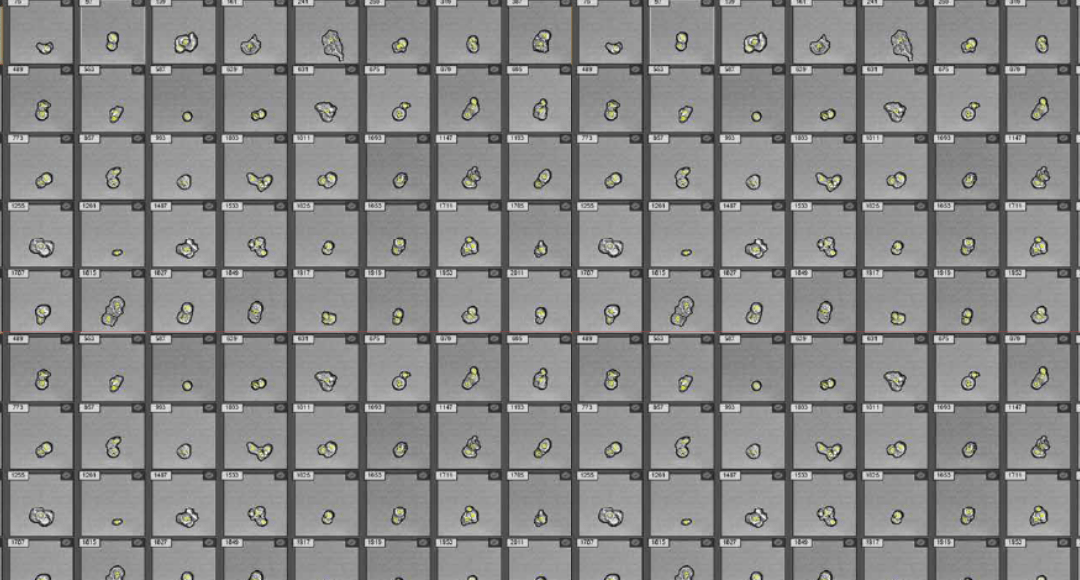
For decades, traditional flow cytometry has been the dominant technique for analyzing single cells within large, heterogeneous populations. However, imaging flow cytometry (IFC) is rapidly becoming more mainstream due to the advantages that it can provide. We spoke...
Guest Authored By: Kelly Lundsten I often think about a concept called the pinball effect. Distilled, it is the knowledge of all of the small changes made by people whose names are remembered, or not, who optimized a process or invented a new chemistry or asked “why”...
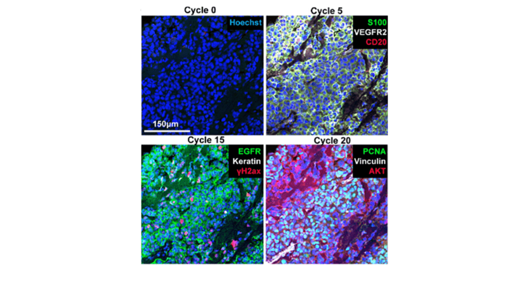
Cover Image: Selected images of a melanoma specimen subjected to 20 cycles of Cyclic IF after cycles 0, 5, 15 and 20. Used without modification under Creative Commons Attribution 4.0 International License from (Lin, 2018) Figure 4G. Spatial biology aims to...
Spectral flow cytometry improves on traditional flow cytometry by capturing the entire emission spectrum of each fluorochrome, rather than isolating specific wavelengths. This enables more precise resolution of overlapping fluorochromes and greater flexibility in...
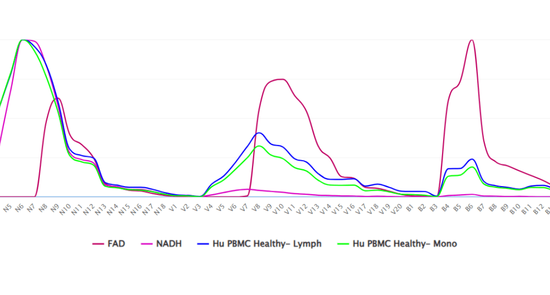
Guest Authored By: Kelly Lundsten Every living organism on earth has evolved to either harness or protect itself from the energy of the sun. Organic molecules capture energy and conduct it along a circuitry of electron pairs shared by double bonds that compose various...
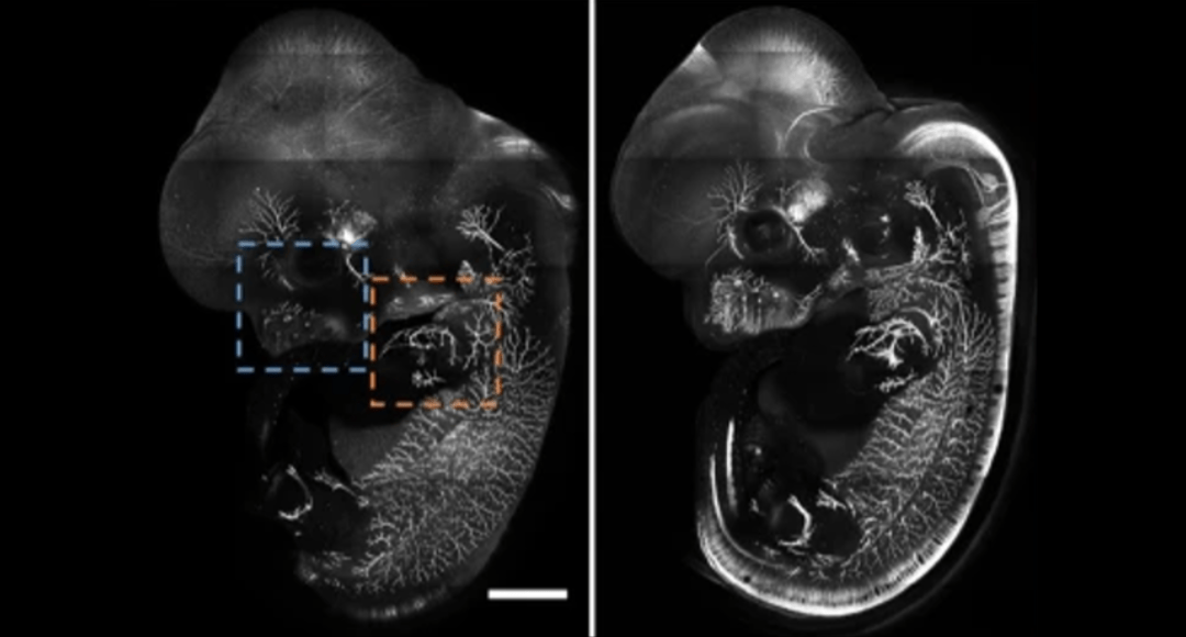
Cover Image: E12.5 whole mouse embryo was immunostained with neurofilament antibody and cleared with RTF. Scale bar, 1000 μm. Used without modification under Creative Commons Attribution 4.0 International License from (Yu T. Z., 2018) Figure 4a. As explained in last...