Sponsored by Slingshot, Written by Nathan Ni
Spectral flow cytometry has proven revolutionary for studying and characterizing cells, giving scientists the power to investigate up to fifty different fluorophores in a single panel. This lets them delve deeper into complex cell populations and rare subsets, illuminating the inner workings underpinning health and disease.1
Spectral unmixing lies at the heart of spectral flow cytometry, and scientists need accurate reference spectral signatures to ensure spectral unmixing validity and consistency. While cell-based reference standards remain ideal, commercially available spectral unmixing options are becoming progressively more cell-like.
Controls for Spectral Unmixing
Conventional flow cytometry uses a dedicated detector for each fluorochrome, and therefore can only investigate as many fluors simultaneously as there are detectors. In contrast, spectral flow cytometry captures the full emission spectrum across multiple detectors. This combined signal is then unmixed to identify distinct spectral signatures corresponding to individual fluorophores.2
As more fluorophores are added to a panel, the more overlap there will be between their respective emission spectra and the harder it becomes to properly distinguish individual signals. Scientists rely on reference spectral signatures to ensure spectral unmixing accuracy.
The most ideal reference control is a single-color spectral signature produced from a fluorophore bound to the cells of interest, as this approach accounts for autofluorescence and other background noise.2 However, this is not always possible, usually due to limited biological material, rare cell populations, or low target marker expression. When cell-derived reference controls are unavailable, researchers turn to synthetic unmixing controls — microparticles that specifically bind fluorochrome-labeled antibodies of interest to generate a cell-like signal during flow cytometry.
A Spectral Unmixing Control that Mimics Cells
Spectral unmixing beads are effective reference controls with several clear advantages: they are easy to obtain commercially and consistently bind a known quantity of antibody.3 However, they are not yet perfect analogs for cells.2,3 SpectraComp® unmixing & compensation controls from Slingshot Biosciences are one example of how scientists are trying to bridge the gap between beads and cells. These synthetic cell mimics use a polymer composition to produce a more cell-like light scatter profile and autofluorescence spectra.
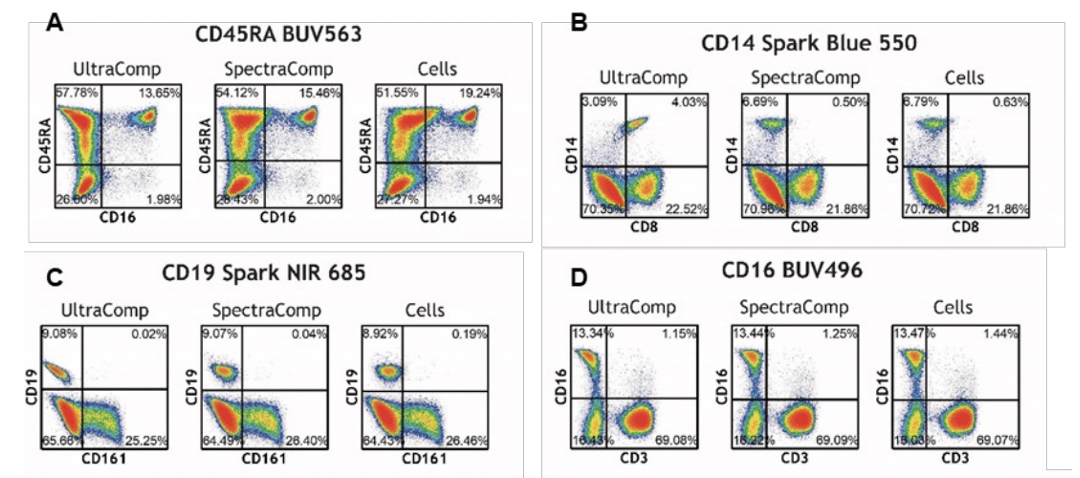
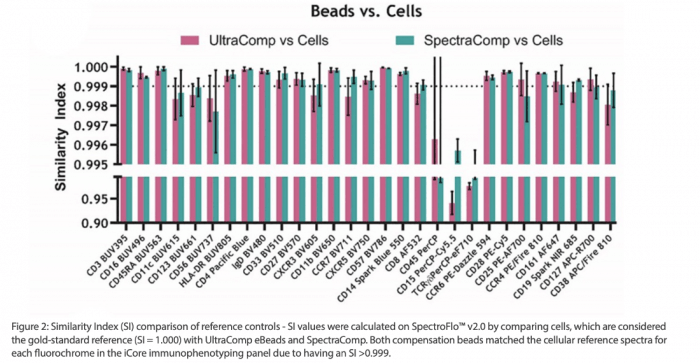
A study by Holly Jensen and Jeong Kim from Bristol Meyers Squibb examined how SpectraComp compared to cell-based reference controls as well as polystyrene beads as part of developing a thirty-color spectral flow cytometry panel for immune biomarker analysis.4 They found that SpectraComp controls showed strong spectral overlap with cell controls for 23 of 30 panel markers, as determined by having a Similarity Index greater than 0.999.
Interestingly, this study noted that SpectraComp reference controls improved unmixing results compared to cell-based controls despite having essentially identical spectral profiles, highlighting that CD45RA BUV563, CD14 Spark Blue 550, and CD19 Spark NIR 685 SpectraComp controls all improved signal for neighboring or correlated markers.4
A Better Control
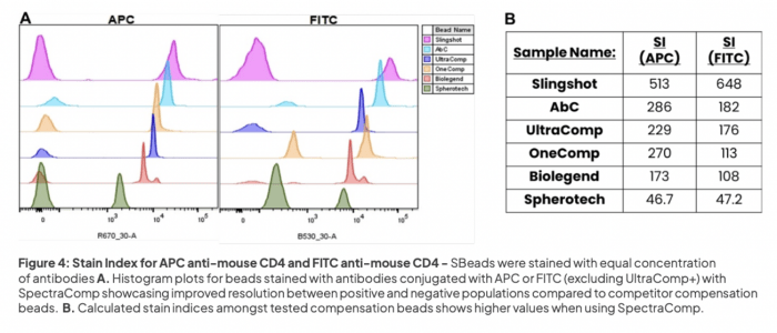
The Jensen and Kim study also found that SpectraComp controls outperformed another commercially available polystyrene unmixing bead. This finding was corroborated by work from Richard Grenfell and his team at the University of Cambridge.5 In their study, Grenfell and his colleagues compared SpectraComp beads with eight other commercially available options in terms of resolution, relative brightness (stain index), event rate consistency and speed, and cost. SpectraComp exhibited the most consistent event rates and the best stain index among the beads tested, achieving the top overall ranking based on all evaluation criteria.
Conclusion
As highlighted by two independent studies, Slingshot Biosciences’ SpectraComp controls are narrowing the gap between bead-based and cell-based controls. SpectraComp is a robust and practical alternative to cell-based controls for compensation and spectral unmixing in most circumstances, especially when working with rare populations where cell numbers are limited. SpectraComp offers improved spectral resolution and reduced errors associated with spectral overlap, providing more accurate compensation and unmixing resulting in higher-quality data.
Supporting your Research
FluoroFinder has developed a suite of tools to help streamline research logistics, including a Flow Cytometry Panel Design tool that simplifies the process of selecting the best fluorophore combinations and our Spectra Viewer, which lets scientists compare the spectral properties of more than 1,000 dyes alongside instrument-specific laser and filter configurations.
* Slingshot Biosciences’ cell mimics are for research use only (RUO) and are not intended for use in clinical or diagnostic applications.
Resources
- Sharma S et al., A practitioner’s view of spectral flow cytometry. Nat Methods. 2024;21(5):740-743. doi: 10.1038/s41592-023-02042-3.
- Mage PL et al., Measurement and prediction of unmixing-dependent spreading in spectral flow cytometry panels. bioRxiv [Preprint]. 2025:2025.04.17.649396. doi: 10.1101/2025.04.17.649396.
- Shevchenko Y et al., Fluorochrome-dependent specific changes in spectral profiles using different compensation beads or primary cells in full spectrum cytometry. Cytometry A. 2024;105(6):458-463. doi: 10.1002/cyto.a.24836.
- Jensen HA, Kim J. iCoreDrop: A robust immune monitoring spectral cytometry assay with six open channels for biomarker flexibility. Cytometry A. 2023;103(5):405-418. doi: 10.1002/cyto.a.24708.
- Grenfell R, et al., “Freaky Flow: The Disappearance of Compensation Beads.” Poster presented at: CYTO 2025, May 31st to June 4th, 2025, Denver CO.


