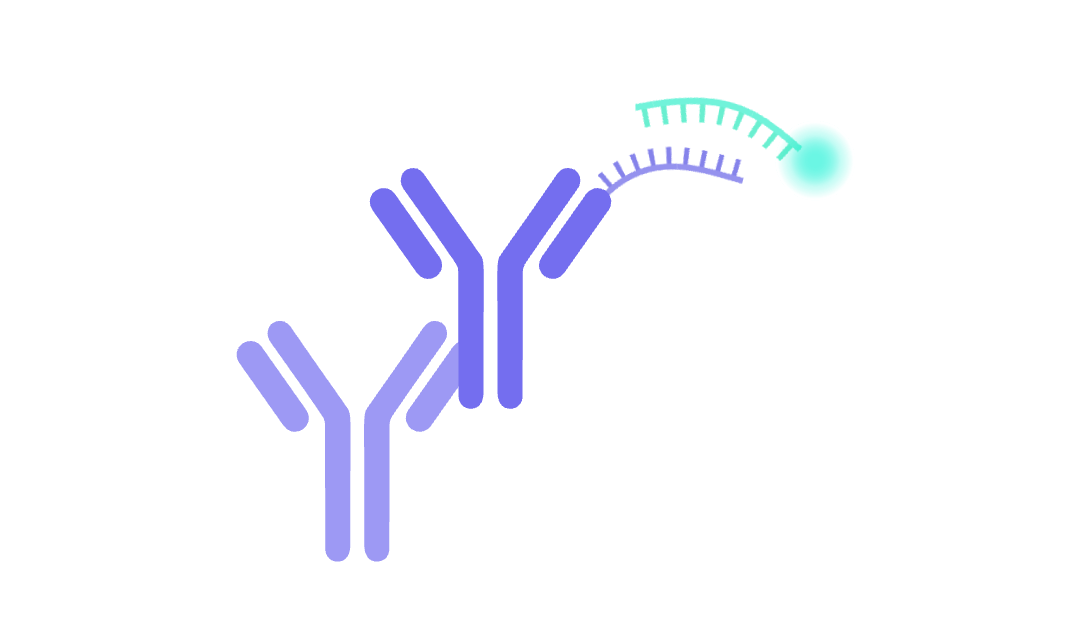
DNA-PAINT (DNA points accumulation for imaging in nanoscale topography) is a super-resolution microscopy technique that exploits the transient binding of fluorescently labeled DNA probes. It has been widely adopted for scientific research owing to its accessibility, although the original method has undergone countless modifications to address various imaging challenges. This article explains the principles of DNA-PAINT and looks at how the technology is evolving.
Introduction to DNA-PAINT
To put DNA-PAINT into context, recall that SRM techniques break through the optical resolution limit in one of two main ways: reducing the size of the point spread function or using single molecule localization. DNA-PAINT falls into the second category, along with methods such as STORM (stochastic optical reconstruction microscopy) and PALM (photoactivated localization microscopy). However, while STORM and PALM rely on photoswitchable fluorescent probes to resolve spatial differences, DNA-PAINT uses fluorescently labeled DNA oligomers.
The original DNA-PAINT method was derived from PAINT, a technique first demonstrated in 2006 by Sharonov and Hochstrasser (1). Using Nile Red, a lipophilic stain that is weakly fluorescent in water but fluoresces brightly in hydrophobic environments, the duo were able to obtain super-resolution images of large unilamellar vesicles through successive cycles of binding, localization, and photobleaching. In 2010, Giannone et al. generalized PAINT to form uPAINT (universal PAINT), which utilizes different fluorophore/ligand combinations for imaging specific membrane proteins on living cells (2).
At around the same time that uPAINT was reported, DNA-PAINT was developed by Jungmann et al., who recognized that the interaction between two complementary DNA strands could provide the basis for a controlled, transient interaction (3). By generating fluorescent DNA oligomers, capable of binding and unbinding from the target strands of DNA origamis, the team were able to demonstrate <30 nm resolution.
Principles of DNA-PAINT
DNA-PAINT uses pairs of complementary DNA strands (~8–10 nucleotides in length) to visualize targets of interest. One strand, known as the docking strand, is typically linked to a target-specific antibody. The other, known as the imager strand or imager probe, is covalently attached to a fluorophore and allowed to diffuse freely in solution. When the two strands hybridize, an increase in fluorescence is observed until the imager strand is released. The duration of each fluorescent ‘blink’ is influenced by the DNA sequence, which determines the off-rate, and the concentration of the imager strand. By capturing multiple blinks over time, researchers can construct a super-resolution image of their sample using SMLM algorithms.
Unlike STORM and PALM, DNA-PAINT is not temporally constrained by photobleaching since the fluorophores are continually replenished by new imager strands. Importantly, DNA-PAINT allows for using standard instrumentation and existing antibody reagents, which are easily conjugated in-house or by an external service provider.
Advancements in DNA-PAINT
A main limitation of the original DNA-PAINT method is that the high background resulting from unbound imager probes limits the imaging speed and throughput. Additionally, it detects just a single target and does not allow for studying proteins inside living cells. To address these constraints, DNA-PAINT has evolved into techniques including the following:
Exchange-PAINT
Developed by Jungmann et al. in 2014, exchange-PAINT enables sequential imaging of multiple targets using only a single dye and a single laser source (4). Initially, the targets of interest are labeled with orthogonal docking strands. The first imager strand is then introduced and an image is captured, prior to washing and addition of the next imager strand. With this approach, Jungmann et al. obtained ten-“color” super-resolution images for DNA structures and four-“color” super-resolution images for fixed HeLa cells.
PRISM
PRISM (probe-based imaging for sequential multiplexing), reported by Guo et al. in 2019, involves simultaneously immunostaining multiple targets with docking strand-conjugated antibodies before sequentially applying imager probes specific to each marker (5). Confocal microscopy is performed using single-stranded locked nucleic acid (ssLNA) imager probes, which bind stably but reversibly to the docking probes, while super-resolution PAINT imaging uses low-affinity versions of the same imager probes to resolve nanometer-scale protein organization. With PRISM, Guo et al. were able to concurrently examine nine synaptic targets.
DNA-PAINT-ERS
DNA-PAINT-ERS, developed by Civitci et al. in 2020, enhances DNA hybridization kinetics and efficiency through the additions of ethylene carbonate (E) to the imaging buffer, sequence repeats (R) to the docking strand, and a spacer (S) between the docking strand and the affinity agent (6). These modifications are applicable to most, if not all, existing docking and imager strand pairs, enabling the integration of DNA-PAINT-ERS with current workflows.
LIVE-PAINT
In LIVE-PAINT (live cell imaging using reversible interactions-PAINT), reversible peptide–protein interactions are responsible for the transient localizations needed for SMLM (7). The method, first demonstrated in 2020 by Oi et al., involves genetically fusing a short peptide to the protein of interest, which is expressed from its endogenous promoter. Additionally, a peptide-binding protein is genetically fused to a fluorescent protein and expressed from an inducible promoter. The small size of the peptide tags enables fluorescent labeling of targets that do not tolerate direct fusion to a fluorescent protein and has been successfully used to track several protein targets inside live S. cerevisiae.
Repeat DNA-PAINT
Repeat DNA-PAINT was reported by Clowsley et al. in 2021 as a method for suppressing background noise, which can be especially problematic when imaging deep in biological tissues (8). By introducing multiple repeat domains into the docking strands, the team were able to significantly reduce the imager probe concentration. This provided a 5-fold reduction in free-imager background and a 10-fold reduction in non-specific events, as well as accelerated data acquisition by approximately 6-10 times.
Fluorogenic DNA-PAINT
The development of fluorogenic DNA-PAINT provided a 26-fold increase in imaging speed over regular DNA-PAINT and made it possible, for the first time, to perform 3D DNA-PAINT imaging without optical sectioning (9). Developed by Chung et al. in 2022, fluorogenic DNA-PAINT uses imager probes that are conjugated to both a fluorophore and a quencher such that they do not fluoresce when free in solution. In addition, the docking strands feature internal mismatches for faster off-rates that accelerate image acquisition. With fluorogenic DNA-PAINT, it is possible to perform simultaneous imaging in separate color channels by using two or more probe combinations.
SUM-PAINT
SUM-PAINT (secondary label-based unlimited multiplexed DNA-PAINT), reported by Unterauer et al. earlier this year, shortens imaging times by decoupling DNA barcoding from the imaging process through the use of transient adaptors (10). First, target-specific antibodies are incubated with secondary nanobodies that each carry a unique DNA barcode. Next, the adaptors (each featuring a speed-optimized docking sequence and a toehold for signal extinction) are hybridized to the respective primary barcodes. Lastly, fluorophore-labeled, speed-optimized imager strands are added for target visualization.
FLASH-PAINT
Like SUM-PAINT, FLASH-PAINT (fluorogenic labeling in conjunction with transient adapter-mediated switching for high throughput DNA-PAINT) uses adaptors to increase the speed of label exchange and expedite image acquisition (11,12). However, with FLASH-PAINT, the adaptor and imager strands are both applied at the same time. Following image capture, the adaptor is removed with an eraser, which stably binds to the adaptor to prevent it from interacting with the docking strand. Using FLASH-PAINT, Schueder et al. have performed applications that include mapping nine proteins in a single mammalian cell and quantifying inter-organelle contacts at 3D super-resolution.
The Future of DNA-PAINT
Besides the methods covered here, many other forms of DNA-PAINT have been (and continue to be) developed. These include techniques such as qPAINT and FRET-PAINT, as described by Ralf Jungmann, and dual-color DNA-PAINT single-particle tracking, which represents a promising method for simultaneous dual-color tracking of proteins on live cells (13,14). As DNA-PAINT continues to evolve, including through its integration with other imaging technologies, super-resolution imaging looks set to become even more powerful.
Supporting Your Research
FluoroFinder has tools to help integrate DNA-PAINT techniques into your microscopy experiment designs. Our Spectra Viewer enables researchers to find compatible fluorophores and reagents with their microscope’s specific laser and filter configurations, and our Dye Directory has a collection of fluorophores from all major suppliers.
References
- https://pubmed.ncbi.nlm.nih.gov/17142314/
- https://pubmed.ncbi.nlm.nih.gov/20713016/
- https://pubmed.ncbi.nlm.nih.gov/20957983/
- https://pubmed.ncbi.nlm.nih.gov/24487583/
- https://pubmed.ncbi.nlm.nih.gov/31558769/
- https://pubmed.ncbi.nlm.nih.gov/32859909/
- https://pubmed.ncbi.nlm.nih.gov/32820217/
- https://pubmed.ncbi.nlm.nih.gov/33479249/
- https://pubmed.ncbi.nlm.nih.gov/35501386/
- https://www.cell.com/cell/fulltext/S0092-8674(24)00248-4
- https://www.cell.com/cell/abstract/S0092-8674(24)00236-8
- https://pubmed.ncbi.nlm.nih.gov/38889688/
- https://pubmed.ncbi.nlm.nih.gov/30544986/
- https://pubmed.ncbi.nlm.nih.gov/37468504/


