Written by Emma Mason
Flow Cytometry Viability Dyes: How to Measure Cell Health
Dead and dying cells can significantly interfere with fluorescence-based techniques such as flow cytometry. Compared to healthy cells, non-viable cells often exhibit increased autofluorescence and greater non-specific antibody binding. As a result, they can generate false-positive signals and reduce the dynamic range of an experiment, ultimately compromising data quality.
To address this challenge, researchers use viability dyes and cell health assays to distinguish live, dead, and functionally compromised cells. These tools not only improve data accuracy but also provide insight into apoptosis, proliferation, and metabolic activity.
Why Cell Viability Matters in Flow Cytometry
Flow cytometry relies on precise fluorescence measurements to identify and characterize cell populations. However, when dead cells are included in the analysis, they can distort results by contributing background signal and non-specific staining.
In practice, excluding non-viable cells is essential for:
- improving signal-to-noise ratio
- increasing reproducibility
- ensuring accurate population gating
For this reason, viability dyes are often one of the first reagents incorporated into a flow cytometry panel.
Traditional Viability Dyes
Traditional viability dyes, which use the loss of membrane integrity as a key indicator for cell death, can only enter cells with compromised plasma membranes. They are divided into three main groups: DNA-binding dyes, protein-binding dyes, and vital dyes. DNA-binding dyes, such as propidium iodide (PI), 7-aminoactinomycin D (7-AAD), and Thermo Fisher Scientific’s SYTOX™ Dyes, emit fluorescence upon intercalating with DNA, enabling non-viable cells to be easily identified. Protein-binding dyes, including BioLegend’s Zombie Dyes, Biotium’s Live-or-Dye™ dyes, and the Image-IT™ DEAD Green™ Viability Stain from Thermo Fisher Scientific, react with primary amines to fluorescently label cellular proteins. In live cells, primary amines are only accessible at the cell surface, resulting in weak fluorescence. But in dead cells, protein-binding dyes can also react with intracellular amines, generating a much higher fluorescent signal. Vital dyes, like alamarBlue®, calcein acetoxymethyl ester (calcein AM), and 5-carboxyfluorescein diacetate acetoxymethyl (5-CFDA) produce fluorescence when acted upon by metabolically functioning cells.
Monitoring Apoptosis
Besides using traditional viability dyes to monitor cell health, it is also common for researchers to investigate apoptotic activity. An established approach uses Annexin V dye conjugates to detect the translocation of phosphatidylserine (PS) from the intracellular to the extracellular side of the plasma membrane. Another widely-used method, known as the terminal deoxynucleotidyl transferase-mediated dUTP nick-end labeling (TUNEL) assay, relies on fluorescently-labeled dUTP to identify apoptosis-induced double-strand DNA (dsDNA) breaks in fixed cells and tissues.
Apoptosis is also monitored by measuring the activity of specific apoptotic proteases with specialized reagents. For example, Biotium’s NucView® Caspase-3 Substrates release a fluorescent DNA-binding dye (emitting either blue, green, or yellow fluorescence) following caspase cleavage. Additionally, apoptosis can be assessed with assays that detect the loss of mitochondrial potential (an early apoptotic event) through changes in fluorescence. These use cationic dyes like JC-1, tetramethylrhodamine methyl ester (TMRE), and Biotium’s MitoView™ 633, which preferentially localize in the matrix of polarized mitochondria.
Tracking Cell Proliferation
Another popular way of evaluating cell viability is to track proliferation. A longstanding method uses carboxyfluorescein succinimidyl ester (CFSE), a membrane-permeant dye that is non-fluorescent until entering viable cells. Here, it is hydrolyzed by cytoplasmic esterases and retained in the cell, such that each cell division reduces the fluorescent signal by half.
To overcome known limitations of traditional CFSE, including leakage from the cell, cytotoxicity, and bleed-through into the PE and PE-TexasRed® channels, several alternatives have been developed. These include Thermo Fisher Scientific’s CellTrace® Violet Cell Proliferation Kit, Bio-Rad’s CytoTrack Cell Proliferation Assays, and Biotium’s ViaFluor® SE Cell Proliferation Kits (available as ViaFluor® 405 SE, ViaFluor® 488 SE, and ViaFluor® 650 SE).
Researchers also have the option to monitor cell proliferation using BioLegend’s Swift-Click™ Green EdU Kit, which is based on the fluorescent detection of a nucleoside analog incorporated into nascent DNA. Fluorescent EdU labeling is catalyzed by Click chemistry, a Nobel Prize-recognized chemical reaction that rapidly links fluorophore-azide molecules to the alkyne group present in EdU in the presence of Cu2+ ions.
Advanced Tools for Cell Health Analysis
To complement the products discussed above, many additional tools have been developed. These include innovative solutions from the following companies:
- BioLegend
BioLegend’s original lineup of Zombie Dyes (Zombie UV™, Zombie Violet™, Zombie Aqua™, Zombie Yellow™, Zombie Green™, Zombie Red™, and Zombie NIR™) now includes five additional products (Zombie UV387™, Zombie YG581™, Zombie R685™, Zombie R718™, and Zombie B550™), increasing flexibility for protein-binding-based discrimination of live and dead cells. Other novel BioLegend products for monitoring viability and cell health include Helix NP™ NIR, a far-red emitting DNA-binding dye, and Apotracker™ Green, which detects the translocation of PS residues to the cell surface in cells undergoing apoptosis.
Discover BioLegend’s extensive selection of Cell Health and Proliferation products.
- Bio-Rad
Included among Bio-Rad’s newer products for assessing cell viability are its range of VivaFix™ Cell Viability Assays, which contain proprietary protein-binding dyes in 7 different colors (VivaFix™ 353/442, VivaFix™ 408/512, VivaFix™ 410/450, VivaFix™ 498/521, VivaFix™ 547/573, VivaFix 583/603™, and VivaFix™ 649/660).
Bio-Rad also offers a selection of 8 different StarBright™ Dye Annexin V conjugates, which allow researchers to detect cells undergoing apoptosis (StarBright™ Blue 675, StarBright™ Blue 765, StarBright™ UltraViolet 400, StarBright™ UltraViolet 795, StarBright™ Violet 440, StarBright™ Violet 515, StarBright™ Violet 790, and StarBright™ Yellow 800).
In addition, Bio-Rad has developed FLICA Caspase Apoptosis Detection Kits for analyzing active caspases using a flow cytometer, fluorescence microscope, or microplate reader. These contain a unique cell-permeable and non-cytotoxic reagent, known as Fluorochrome Inhibitor of Caspases (FLICA), which consists of a caspase inhibitor sequence linked to a fluorescent probe and generates a fluorescent signal that corresponds directly to the amount of active caspase.
- Biotium
Besides the products already mentioned above, Biotium offers many other innovative solutions to monitor viability and cell health. Notably, its amine-reactive Live-or-Dye™ Fixable Viability Stains now come in 18 different colors, including spectrally unique dyes for spectral flow cytometry.
See Biotium’s Cell Viability Blog for additional information.
- Promega
For researchers wishing to evaluate their samples in 96- or 384-well microplates, Promega’s CellTiter-Blue® Cell Viability Assay harnesses the metabolic capacity of living cells to convert a redox dye (resazurin) into a fluorescent end product (resorufin). The homogeneous add-incubate-measure protocol minimizes handling steps and allows for direct addition to serum-supplemented cell cultures without washing or lysis, while the non-toxic reagent formulation enables extended incubations and multiple measurements from the same sample population.
- Proteintech
Proteintech’s Phantom Dyes, which are amine-reactive, further increase the number of options for experimental design. Phantom Dyes are available in 7 different colors (Phantom Dye Red 710, Red 780, UV 450, Violet 450, Violet 510, Violet 540, and Blue 516) and can be used with either live or fixed cells. Additionally, Phantom Dyes can serve as a compensation control when paired with amine-reactive beads.
Find out more about Phantom Dyes and other common methods for assessing cell viability in this Proteintech Blog.
- Revvity
Revvity offers a suite of PhenoVue™ Reagents and Kits for visualizing key reporters of cell health, including measures for viability, apoptosis, mitochondrial membrane potential, oxidative stress, and indicators for calcium and hypoxia. Products include the PhenoVue™ Live/Dead Cell Viability Assay Kit, which uses calcein AM and PI to simultaneously identify live and dead cells; the PhenoVue™ 505 Live Cell Caspase-3/7 Activity Stain, which is a no-wash, cell-permeable fluorogenic dye for visualizing apoptotic cells; and PhenoVue Mitochondrial Stains, which are available in 3 different colors (PhenoVue™ 551, PhenoVue™ 578, and PhenoVue™ 641. Additionally, Revvity’s PhenoVue™ DNA Damage Response Staining Kit contains PhenoVue™ Hoechst 33342 for precise nuclear staining and specific antibodies targeting γ-H2AX and 53BP1 to enable robust, quantitative assessment of DNA damage pathways.
- Thermo Fisher Scientific
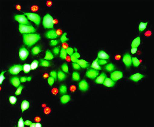
In addition to SYTOX Dyes, the Image-IT™ DEAD Green™ Viability Stain, and various CellTrace products, Thermo Fisher Scientific has developed many other solutions for monitoring viability and cell health. These include a broad array of LIVE/DEAD Fixable Viability Dyes, which offer 2- or 3-color discrimination of live from dead cell populations based on membrane integrity, esterase activity, metabolic activity, or structural segmentation. For example, the LIVE/DEAD™ Viability/Cytotoxicity Kit simultaneously stains live cells green using calcein AM and dead cells red using ethidium homodimer-1 (EthD-1). Other products in Thermo Fisher Scientific’s extensive portfolio include the PrestoBlue™ and PrestoBlue™ HS Cell Viability Reagents, which identify viable cells based on the reduction of resazurin to fluorescent resorufin, and the Vybrant™ Cell Metabolic Assay Kit, which uses C12-resazurin in place of resazurin for improved cellular retention.
Visit Thermo Fisher Scientific’s Selection Guide For Cell Viability Assays and Live Cell Imaging Resource Page
Conclusion
Flow cytometry viability dyes are essential for ensuring accurate and reproducible data. By distinguishing live, dead, and functionally compromised cells, these tools improve experimental reliability and enable more precise interpretation of results.
Ultimately, no single assay can capture every aspect of cell health. Instead, researchers must select the appropriate combination of dyes and probes based on their experimental goals. As new technologies continue to emerge, the ability to measure cell viability and function will become increasingly sophisticated, supporting more complex and informative biological analyses.
To support panel design and reagent selection, researchers can explore FluoroFinder’s Dye Directory to search and compare hundreds of fluorophores and viability dyes. The Spectra Viewer also enables users to visualize spectral properties, assess overlap, and optimize dye combinations for their specific instrument configuration.


