Intracellular Flow Cytometry Fundamentals
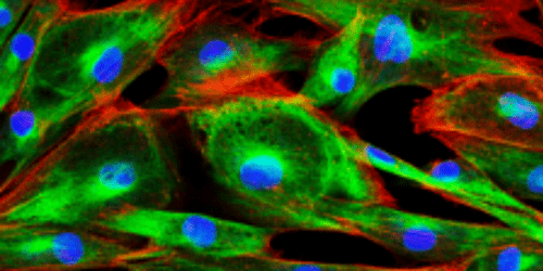
Proteins produced by cells get shuttled around and end up in a variety of locations, whether they are embedded on the cell surface, contained internally within the cell, or secreted outside of the cell. Intracellular flow cytometry refers to the process of staining and analyzing proteins localized inside a cell. In this newsletter, we will go over the basics of intracellular flow cytometry, some important considerations, and how FluoroFinder can help you set up your intracellular flow cytometry experiments. While extracellular staining is relatively straightforward, there are extra steps and considerations required when preparing cells for intracellular staining. Living cell membranes naturally block out most of the fluorescent antibody staining reagents that are useful for intracellular cytometric analysis. Extra preparation steps are necessary to modify the cell to allow the entry of these staining reagents. First, a fixation step stabilizes the cellular structure, then a permeabilization step creates pores in the cell membrane that allow staining reagents to enter the cell.
Cell Fixation
Fixation refers to the stabilization of the cellular structure prior to permeabilizing the cellular membrane and can be accomplished with a few different reagents. This step essentially freezes the cells in place which preserves the overall structure but also kills the cells. While this is not ideal for studying the cell’s ongoing natural processes, freezing cells in place like this allows researchers to evaluate a snapshot of the cell’s state at a specific point in time. This process is necessary because without fixation the cells would rupture when permeabilized, preventing any subsequent analysis. To fix cells, aldehydes including formaldehyde (also known as formalin) may be used. Formaldehyde crosslinks proteins and nucleic acids to preserve the cell’s structure. Overall, Formaldehyde works well but can cause cell surface protrusions known as blebs among other alterations. Formaldehyde is also a carcinogen and is hazardous to work with, so this can direct researchers to other non-toxic methods, a one-step protocol where aldehydes are not used for initial fixation can adversely alter cell structure to the detriment of experimental aims. There are disadvantages when treating cells with any of these harsh chemicals, but the aims of your experiment will let you know which detrimental side-effects your experiment can tolerate while still gathering all of your important data points. Additionally, the exact fixation protocol can vary depending on the type of protein targeted for analysis. For example, transport inhibitors can be used to contain secreted cytokines within the cell for intracellular analysis. Consider all these factors prior to choosing a fixation protocol.
Permeabilization
The permeabilization process makes the cell membrane more porous so that larger molecules such as antibody-fluorochrome conjugates, which would normally be excluded from live cells, may enter the cell to access their intracellular targets. This can be achieved by using organic solvents such as methanol and ethanol, and detergents such as Triton X-100 or Tween 20, which remove lipids and cholesterol groups from the membrane. Consider the strength of the permeabilization agent with your experimental aims; stronger more concentrated agents may be needed when permeabilizing the nuclear membrane in addition to the outer cell membrane when staining nuclear targets.
Intracellular and Extracellular Staining Combined
What if you want to stain both intracellular and extracellular targets in the same experiment? There are multiple ways to do this, but often the best technique is to stain the extracellular proteins, fix and permeabilize the cells, then stain intracellular proteins. One reason for this is that you may want to evaluate a set of markers as they exist extracellularly only, so you do not want your extracellular staining reagents to have access to the proteins on the inside of the cells. Another reason is that the fixation and permeabilization process alters the external cellular structure and may negatively impact how extracellular markers are presented. When using this type of protocol, be sure to research fluorochrome characteristics and look for fluorochromes that are stable when treated with fixatives. Fixation chemicals are harsh and may negatively impact the brightness of fluorochromes in the experiment, particularly with some tandem dyes.
Need to do some research on which fluorochromes are right for your intracellular experiments? FluoroFinder’s new comprehensive Fluorescent Dye Directory provides researchers with a single place to access information on all suppliers’ fluorescent dyes. This is a recent addition to FluoroFinder.com that can help researchers discover which fluorescent dye is suitable for their experiment. For example, is a fluorescent dye susceptible to signal degradation when treated with fixatives? New fluorochromes are routinely added by suppliers so be sure to keep an eye on the latest dyes in this growing library.


