Written by Kelly Lundsten
Early in my career, I made the jump from neuroscience to immunology when I joined a biotech company that manufactured antibodies for flow cytometry. At the time, customers would send in lists of antigens and ask for help designing 17-color panels, which felt incredibly ambitious then. My own learning curve was steep. To build useful panels, I had to understand not just the markers themselves, but why researchers were using them and how they planned to analyze the data.
I kept wondering: why isn’t there a central resource that explains which markers are needed to define different cell subsets? Antibody vendors sometimes offered posters or website summaries listing relevant antigens, but there was little practical guidance on how to combine markers into a robust assay. How markers and distribution patterns were described in the literature did not make things much easier. Marker combinations varied widely, gating logic was inconsistent, and shorthand terms like hi, lo, +, ++, and +/- were used without much standardization.
That gap in the field stayed with me, so when I joined FluoroFinder I was excited by the chance to create educational content that could genuinely help researchers design better panels, whether they were just getting started or already highly experienced. I spent a long time reading through hundreds of papers, looking at marker combinations, functional validation, and disease-specific applications to identify areas of consensus across the field. What became clear was that our understanding of cell identity is constantly evolving. Marker relevance depends on context, and factors such as sample preparation, age, disease state, clone choice, and protocol can all affect how confidently a population can be defined.
Out of that literature survey came the Cell Types Tool.
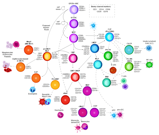
The Cell Types Tool is an interactive resource built around sequential marker strings that reflect our current understanding of how different cell subsets are defined. It currently includes six pages covering human and mouse immune cell subsets, B and T cell maturation, T cell subsets, and human hematopoiesis. Each page provides an overview of subset function and distribution, recommended markers, additional useful markers to consider, and a curated list of key citations.
The interactive image gives users a visual map of how cell subsets relate to one another. Clicking on a subset opens additional descriptive and marker information, and the marker combinations follow a sequential logic as you move along the arrows toward more specific populations. Users can also click directly on markers to explore available clones and products in FluoroFinder’s exhaustive antibody search database. Beneath the image, each page includes a recommended gating strategy. In some cases, there are representative experimental flow plots; in others, there is a schematic version of the strategy until real data can be added. One important difference from more static online resources is that the Cell Types Tool is updated regularly as new papers are published and as researchers provide feedback. It is designed to be a living resource.
When the Cell Types Tool launched in 2024, I did not realize it would also intersect with a broader effort around cell population nomenclature called the SOULCAP Foundation, or Standardized Ontology Unique Labeling of Cell Annotation of Populations.
That effort raises a deceptively simple question: what does it mean to name a cell subset? We often rely on familiar labels like Regulatory T cell or Treg, but those names can hide a lot of ambiguity. Is a Treg defined by a common name alone? By a marker string such as CD45+ CD3+ CD4+ CD8- CD127- CD25hi? Or by CD3+ CD4+ FoxP3+? In the literature, both marker combinations may be described as Tregs, even though they do not necessarily identify identical cell populations. As a result, events captured in the final subset gate are not always the same. Add vague expression terms like high, low, or ++, and the problem becomes even more difficult for downstream interpretation and reproducibility.
There is also an ontological side to the problem. Tregs are immunosuppressive T cells, but the term may also refer to cells from a particular species, tissue, activation state, or developmental context. To help standardize these relationships, the Cell Ontology working group created the Ontology Lookup Search (OLS), an online resource that organizes cell types hierarchically and assigns them unique identifiers known as CL IDs. Over time, these entries will incorporate SOULCAP-recommended marker combinations, providing practical guidance for harmonized panel design and more consistent annotation in single-cell cytometry experiments.
For true standardization, though, consensus terminology is only part of the solution. Annotation also needs to become automated within a bioinformatics framework that can be incorporated into commonly used analysis platforms. One idea being explored is the use of standardized pixel maps overlaid on flow cytometry plots to align cell populations spatially. In this approach, each cluster or population would have a defined density center that supports automated gating, and each event within that population could be assigned spatial coordinates as part of its identity.
Even with variability in instrument sensitivity, reagent brightness, and staining resolution, adding spatial structure to analysis could allow unknown query datasets to be overlaid onto an internal gold-standard dataset generated under matched conditions. Those datasets could then be rasterized and aligned, enabling automated identification and annotation of events. For this to work consistently, however, researchers would need to adopt an additional control that standardizes data scaling. Right now, instrument manufacturers vary widely in how signal intensity is scaled, which makes cross-platform comparison difficult. A more uniform system, potentially supported by scaling standard beads such as MicroCal beads from Cellarcus, could make automated spatial annotation much more reliable.
Looking ahead, broader support from organizations such as IUIS, ICCS, ESSCA, ISAC, and FOCIS, along with their affiliated journals and publications, will help move these standards from aspiration to expectation. If reporting standards become more consistent, that would contribute meaningfully to addressing the reproducibility problems that continue to affect experimental science.
Artificial intelligence will almost certainly play a growing role in scientific discovery. But AI is only as useful as the data it is built on. Without consistent datasets and standardized ways of describing cell populations, even the best algorithms will struggle to generate reliable insights. It is a big challenge, but it is one worth starting now. FluoroFinder is excited to support the efforts of SOULCAP through the maintenance of the Cell Types Tool, an important piece of the strategy towards automated cell annotation in flow cytometry.