Written by Emma Mason
When Benjamin Franklin coined the phrase “by failing to prepare, you are preparing to fail”, he certainly wasn’t referring to flow cytometry, which wouldn’t be invented for another 200+ years. However, the expression is apt—good sample prep is the foundational step of any flow cytometry experiment and it’s critically important that you get it right. We spoke with Amber Miller, Senior Manager for Antibody Services at Bethyl Laboratories, a Fortis® Life Sciences company, who shared practical flow cytometry sample prep tips inspired by her Ph.D. experience working with mouse gut associated lymphoid tissue and by her current work assessing antibody functionality for use in flow cytometry.
Key Takeaways:
-
Flow cytometry sample preparation is critical for generating accurate, reproducible data.
-
Harvesting methods vary by sample type, including suspension cultures, adherent cells, blood, cryopreserved samples, and solid tissues.
-
Maintaining a single-cell suspension and preserving viability help prevent clogs and improve data quality.
-
Fixation, permeabilization, and background reduction steps should be optimized for each experiment and panel.
-
Antibody and fluorophore selection—along with careful panel design—are essential for minimizing spectral overlap and ensuring reliable results.
How to Harvest Cells for Flow Cytometry (by Sample Type)
Broadly speaking, there are five main sources of cells for flow cytometry—suspension cultures, adherent cultures, cryopreserved cells, whole blood, and solid tissues—all of which differ in terms of how easily they can be harvested.
Suspension Cultures
Suspension cultures are the most straightforward, simply requiring centrifugation to pellet the cells, washing in phosphate buffered saline (PBS) to remove the culture medium, and resuspension in a suitable volume of staining buffer to obtain the desired cell concentration (typically 105 – 107 cells/ml).
Tip: use ice cold staining buffer to preserve cellular epitopes
Adherent Cultures
Adherent cultures require several additional processing steps in order to detach the cells from the culture vessel. These include either gently scraping the cells from the culture surface using a sterile cell scraper or adding a dissociation buffer. There are two main classes of dissociation buffers, one containing enzymes like trypsin and the other being enzyme-free and instead containing chelating agents that bind the calcium and magnesium required for cell adhesion (e.g., Gibco™ Cell Dissociation Buffer from Thermo Fisher Scientific). The single-cell suspension can then be prepared as described above.
Tip: if you plan to detect cell surface proteins, consider avoiding trypsin to better preserve epitopes for antibody binding
Cryopreserved Cells
Cryopreserved cells may either be used immediately or allowed to recover in culture medium prior to harvesting. In either scenario, it is essential that the cells are thawed rapidly in a 37°C water bath and that the freezing medium is removed as soon as possible to prevent any cytotoxic effects from DMSO.
Tip: after thawing, transfer the cells to a chilled centrifuge tube and add ice cold culture medium drop by drop until the cells are diluted 10X
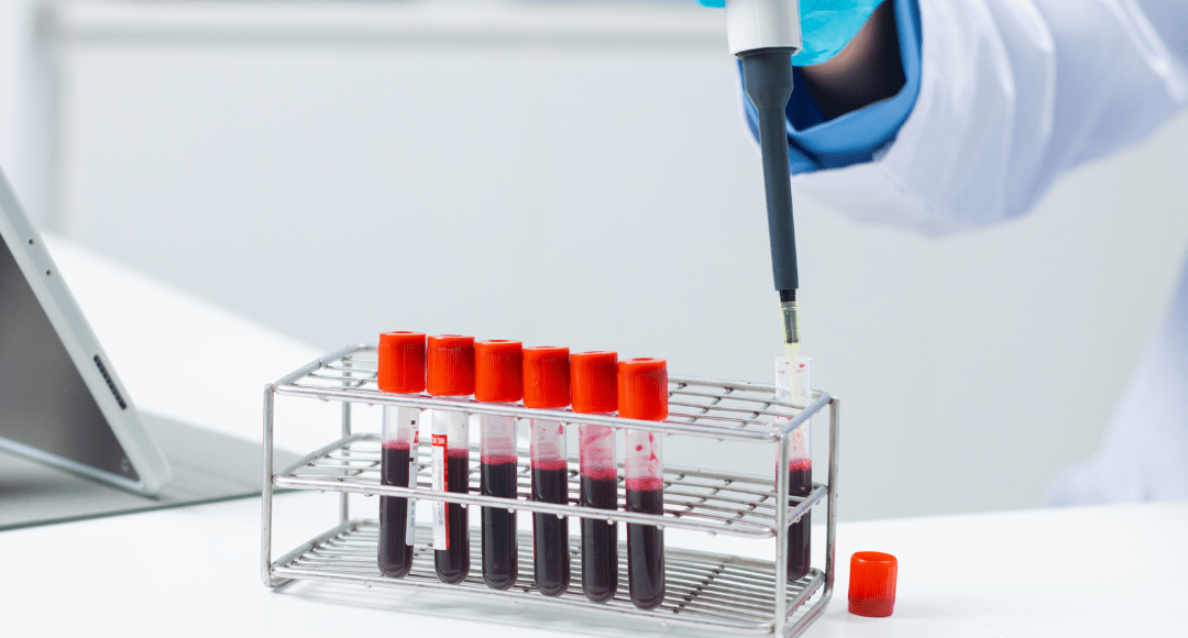
Whole Blood
Whole blood is more challenging to work with, generally requiring that a multi-step protocol be used for isolating peripheral blood mononuclear cells (PBMCs). A common approach involves diluting the blood 1:1 with PBS and layering it over a gradient medium (e.g., Ficoll or Histopaque®) before aspirating the PBMCs from the serum/medium interface for washing and resuspension. Alternatively, whole blood separation systems such as BD Vacutainer® CPT™ Mononuclear Cell Preparation Tubes can be used to reduce the number of protocol steps and minimize experimental variability.
Tip: blood collection tubes typically contain EDTA, citrate, or heparin as an anti-coagulant; always check that the anti-coagulant is compatible with your downstream application1
Solid Tissues
Solid tissues necessitate unique processing steps, including mechanical and/or enzymatic disaggregation to disrupt the tissue structure and treatment with Erythrolyse Red Blood Cell Lysing Buffer or a similar reagent to remove contaminating red blood cells (RBCs). When handling these types of samples, understanding the tissue architecture is key to maximize the yield of viable cells. For example, while a single-cell suspension may be prepared from bone marrow using mechanical disruption alone, skin tissue may require sequential treatment with dispase (to separate the epidermis from the dermis), collagenase (to disrupt the extracellular matrix), and trypsin (to release individual cells).
Tip: enzyme dissociation can modify cell surface proteins, so be sure to validate the process for your flow cytometry panel
Cell Counting and Viability Assessment for Flow Cytometry
Once you’ve produced a single-cell suspension, the next step is to perform a cell count and check the viability of your sample. Traditionally, this would involve staining an aliquot of the sample with Trypan Blue and using a hemocytometer to manually count live and dead cells. However, because this method is both time-consuming and subjective, automated cell counters are now used more frequently. Available options include the Vi-CELL BLU Cell Viability Analyzer from Beckman Coulter Life Sciences, the Invitrogen Countess 3 Automated Cell Counter from Thermo Fisher Scientific, and the LUNA-FX7™ Automated Cell Counter from Logos Biosystems.
Cell counting and viability measurements can also be performed using a flow cytometer. Reagents such as Precision Count Beads™ from BioLegend and CountBright Plus Absolute Counting Beads from Thermo Fisher Scientific enable researchers to determine absolute cell numbers, while products including the BD™ Cell Viability Kit with BD Liquid Counting Beads allow for checking both the cell concentration and viability prior to staining for flow cytometric analysis. Factors to consider when deciding which method to use include how many samples you intend to handle and what instrumentation is available in your lab.
Preventing Cell Clumping in Flow Cytometry Samples
Ensuring that your samples are free of clumps is essential to prevent instrument clogs, maintain a uniform sample stream, and improve data quality. It is recommended that you pass samples through a cell strainer prior to staining, and that you also consider repeating this process ahead of sample acquisition. Cell strainers are available from suppliers including Corning® Life Sciences and Miltenyi Biotec and come in a range of mesh sizes to suit different applications. Other ways to avoid clumping include adding EDTA to the resuspension buffer to chelate Ca2+/Mg2+, which can promote aggregation, and treating samples with DNAse to digest free DNA (released from dead or dying cells) that can otherwise bind cells together.
Tip: if cells are pelleted too hard, they can clump, so be sure to set the speed of your centrifuge correctly
Fixation and Permeabilization for Intracellular Flow Cytometry
While some flow cytometry experiments use live cells, others require that samples be fixed and/or permeabilized. To avoid misinterpretation of results, fixation and permeabilization should be carefully optimized. A key point to note is that protein-based fluorophores like phycoerythrin (PE) and allophycocyanin (APC) are denatured by alcohol-based organic solvents, making them a poor choice when using these types of reagents. Researchers should also be aware that aldehyde-based fixation can generate autofluorescent by-products, meaning that fluorophores with longer emission wavelengths (>550 nm) are often preferred for staining formaldehyde-fixed samples. When staining for both extracellular and intracellular targets, performing iterative protocol optimization with different types of fixatives and permeabilization solutions is recommended.
Tip: read our Guide to Fixation and Permeabilization for further information on these protocol steps
Reducing Background and Autofluorescence in Flow Cytometry
Sources of unwanted background signal include non-specific antibody binding interactions, antibody capture by Fc receptors on immune cells, and autofluorescence, all of which have the potential to generate false positive results. Non-specific antibody binding is typically blocked using reagents such as bovine serum albumin (BSA) or normal animal serums, while Fc receptor binding can be addressed with products such as Human Fc Receptor Blocking Solution from Cell Signaling Technology or Mouse Seroblock FcR from Bio-Rad, or by blocking with the addition of an Fc fragment such as Fortis Life Sciences’ Purified Human IgG-Fc Fragment normal serum. Ways of reducing autofluorescence include using alcohol-based fixatives in place of aldehydes, ensuring that red blood cells are eliminated from samples, and implementing measures to limit the presence of dead cells and debris. The use of appropriate controls is essential to accurately account for unwanted background signal.
Tip: Learn more about autofluorescence and how to reduce it here
Supporting Your Research – Antibody Selection and Panel Design for Flow Cytometry
Selecting compatible antibodies and fluorophores is one of the most complex steps in any flow cytometry experiment—especially as panel size increases and spectral overlap becomes more difficult to manage. Researchers must balance marker expression levels, fluorophore brightness, spillover, and instrument configuration to generate reliable data.
FluoroFinder helps streamline these decisions by enabling researchers to evaluate antibody and fluorophore compatibility before running an experiment. Use Antibody Search to identify reagents validated for flow cytometry, then explore the optical properties and spectral profiles of more than a thousand fluorophores in the Fluorescent Dye Database. The Spectra Viewer allows you to assess dye performance based on your instrument’s lasers and filters, while the Panel Builder helps you design and refine multicolor panels using reagents from 85+ suppliers in a single workspace.
By centralizing panel design considerations—reagent validation, spectral compatibility, and instrument constraints—these tools help reduce trial-and-error and support more confident experimental planning.
References:
- Diks AM, Bonroy C, Teodosio C, et al. Impact of blood storage and sample handling on quality of high dimensional flow cytometric data in multicenter clinical research. J Immunol Methods. 2019;475:112616. https://www.sciencedirect.com/science/article/pii/S0022175919301140


