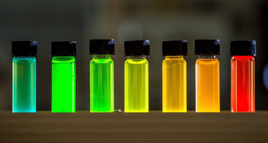
So many new fluorophores have been released in recent years, it can be hard to keep track of their similarities or novelty. Sometimes, a company will create a new brand name for a fluorescent chemistry that is already commonly used, which can make sifting through the relevance of new products a challenge. Let’s break them down by type of chemistry to group fluorophore families with similar characteristics, along with the companies and brand names that fall into these different categories.
Organic Fluorophores
Simple organic fluorophores are the building blocks of all fluorescence found in nature from aromatic amino acids like tryptophan and tyrosine to metabolic products like NADH and FAD to both soluble and insoluble antioxidants like riboflavin and vitamin E (Tocopherol). Common structure of all organic fluorophores is a rigid, conjugated polyaromatic hydrocarbon structure that in its microenvironment, whether an aqueous solution, a lipid membrane or an acidic organelle, it is capable of absorbing energy of a specific wavelength, jumping to an excited state where it can resonate that energy efficiently before emitting it as a photon of light.
These molecules are small, typically less than 2kD and they can be modified to be pH and solvent stable and resist ROS-mediated oxidative degradation like photobleaching. Finally, when they are conjugated to an antibody or other biomolecule, the ratio of fluorophore to protein will reflect the size of both the biomolecule and the fluorophore but is generally 3-6 fluorophore per antibody. This includes their use as the acceptor fluorophore in a FRET tandem with PerCP, PE or APC. Every chemistry serves a special function in an assay and although each has strengths and weaknesses, sometimes even a weakness can be a strength. For example, this family is small and therefore has limited brightness, but this weakness can be used advantageously for fixable viability reagents.
Examples of brand names associated with this fluorophore family includes:
| Brand Names | Vendor | ||
| Cyanine, FITC, Coumarin, TAMRA, ROX, Rhodamine | Off-Trademark | ||
| Brand Names | Vendor | Brand Names | Vendor |
| iFluor®, XFD™ | AAT-Bioquest | cFluor*, Ghost | Cytek |
| HiLyte™ | Anaspec | DyLight™, DY | Dyomics |
| ATTO | ATTO-Tec | Vio®, Vio®Bright, Viobility™ | Miltenyi |
| V450/V500, FVS | BD Biosciences | Janelia Fluor® | Promega |
| ViaKrome, Krome | Beckman Coulter | CoraFluor™ | R&D Systems |
| Spark, Zombie | Biolegend | Seta, SeTau | SETA BioMedicals |
| CF®, Live-or-Dye™ | Biotium | eFluor™, LIVE/DEAD™, Alexa Fluor™, Pacific Blue™, Bodipy™ | Thermo Fisher |
*Indicates exceptions to this brand
Chromoproteins
Protein-based fluorophores, also called chromoproteins, are naturally produced by algae, cyanobacteria, jellyfish, sea anemones, slugs and corals in the form of fluorescent proteins (FP), R-phycoerythrin (R-PE) and allophycocyanin (APC). The majority of the mass of these structures is a non-fluorescent protein scaffolding which embed small chemical chromophores. When a protein is properly folded, these chromophores become fluorescent, also called fluorogenic, due to the structural rigidity created by the protein microenvironment. Thus, the fluorescence of proteins will be very sensitive to anything that can denature, degrade or digest the protein, like any alcohol in a fix and perm buffer.
Fluorescent proteins (FPs) can be monomeric or polymerize together into dimers, etc. Polymeric FPs can be a hazard when using them for protein localization, in the event they are binding to each other. However, polymeric FPs can also be brighter, shift spectrally to a longer wavelength emission and be useful as a functional probe due to spectral changes that can happen upon aggregation.
R-phycoerythrin (R-PE), peridinin-chlorophyll complex (PerCP) and allophycocyanin (APC) are all commonly used reagents in flow cytometry. PE and APC are very large macromolecules with multiple chromophores generally called phycobilins embedded on different subunits, for example PE is 240kD with 24-30 embedded phycobilins and APC is 105kD with 6 phycocyanobilins. PerCP is 30kD with 8 perdinin complexed to 2 chlorophyl molecules and is much dimmer than either PE or APC and much more photo labile. PE and APC make excellent donor molecules coupled to simple organic dyes in a FRET tandem and with the exception of the Cytek, these producs will be named first as PerCP, PE or APC and then the brand name of the simple organic fluorophore to which it is coupled.
Inorganic Nanocrystals
To increase the number of fluorescent parameters flow cytometry, inorganic nanocrystals were introduced as antibody conjugates around 2005. Generally, inorganic nanocrystals will consist of a metallic core like Cadmium Selenide or Cadmium Telluride encased by a thin coating like Zinc Sulfide to seal the core, a technology which won the Nobel Prize in Chemistry 2023. Others may be composed of colloidal aggregations of metal particles. However, a nanocrystal needs to be functionalized to covalently conjugate a biomolecule or antibody, which is a challenge for a metallic particle. Typically, the particle will be coated in either a spray or “net” of lipids or an amphiphilic polymer to ensure even surface coverage.
The overall size of the particle and the hydrophobic surface can cause some issues with intracellular penetration and reagent aggregation respectively. Despite these challenges, nanoparticles have some unique beneficial properties. Because they are inorganic, they do not succumb to photobleaching like organic fluorophores. Their core can be oxidized under intense direct laser penetration, but not nearly at the same rate as the aforementioned simple organic and protein-based fluorophores. These nanocrystals are often used in solar technologies due to their efficient absorption/excitation at the shortest wavelengths, like UV and 405nm lasers. And finally, they can exhibit tight emission profiles with less spectral spillover into neighboring channels.
ThermoFisher offers QDot nanocrystals and CoreQuantum Technologies offers MultiDots within this family. Also of interest are CoreQuantum’s MagDots which incorporate superparamagnetic iron oxide nanoparticles called SPIONs that enables both fluorescent detection and cell separation. More information can be found on applications for MultiDots and MagDots in our on-demand webinar library.
Multimers and Polymers
Organic multimers and polymers were developed to replace and expand QDot nanocrystal applications in flow cytometry. The Horizon™ Brilliant fluorescent polymers from BD Biosciences, the SuperNova dyes from Beckman Coulter, the SuperBright polymers from Thermo Fisher, and some of the VioBright Dyes from Miltenyi are of similar structure and utility off the UV and 405nm laser lines in flow cytometry.
Although they are smaller, Brilliant Violet 421 and Brilliant Ultraviolet 395 have similar extinction coefficients (EC) and brightness to PE and APC respectively. High EC values make them excellent donor molecules in a FRET tandem, creating an array of spectrally unique emission profiles. Because they are synthetic organic chemical structures with no protein component, they are also very stable to temperature, solvent and fixatives. However, this chemistry is known to have significant unwanted non-specific binding characteristics, specifically to itself, and a staining buffer is required to prevent this.
From Bio-Rad, an alternative polymer dot called StarBright reagents, overcomes some of these non-specific binding issues and even more importantly offers polymer dots, or pdots, capable of excitation off the 488nm, 561nm and 633nm lasers.
Another advancement in multimer dye technology is the RealBlue and RealYellow fluorophores from BD Biosciences. RealBlue and RealYellow fluorophores are smaller than the traditional polymer chemistry with better intracellular and intranuclear permeability, and a very high brightness.
As the competition grows around this chemistry, we are sure to see even more improvement in the quality, stability and reliability of multimers and polymers.
Supporting Your Research
Our previous article on the Expansion of Fluorophores for Spectral Flow Cytometry may be of interest to you for additional reading on fluorophores. This resource also provides information on DNA backbone polymers that were not described here.
You can use the Dye Directory to search our comprehensive list of fluorophores with spectra and photophysical characteristics and a view of spectrally similar fluorophore alternatives for comparison.


