High viabilities, well-preserved antigens, and minimal clumping underpin flow cytometry success
Unlike bulk population analysis techniques such as ELISA and Western blot, flow cytometry provides information about individual cells. Although microscopy does this as well, microscopy has a much lower throughput than flow cytometry, risking rare cell types being overlooked. For flow cytometry to deliver accurate results, it is critical for the sample material to be in the form of a single-cell suspension, in which individual cells are free floating in suspension as opposed to being clumped together. However, this involves far more than just pipetting samples up and down a few times before introducing them into the flow cytometer. Instead, sample preparation must be carefully optimized for the system in question to ensure that cells retain high viabilities, display intact antigens, and are unlikely to clump together.
Why is having a single-cell suspension so important?
To fully appreciate the importance of having a single-cell suspension for flow cytometry, it’s worth taking a moment to recall how a flow cytometer works. First, the sample is introduced into the fluidics system, which directs the cells through the flow cytometer in a single file. As each cell crosses the instrument’s interrogation point, it scatters the laser lines that are focused there and, at the same time, emits fluorescence from any cell-bound fluorophores. The resultant signals are then directed by a series of lenses, mirrors, and filters towards the flow cytometer’s detectors, which capture light of a specific spectral band and convert it into a voltage pulse (known as an event) that can subsequently be analyzed.
Now, consider that an event correlates with the progress of each cell through the interrogation point, whereby the voltage rises as the cell enters the interrogation point and returns to baseline levels as it leaves. Each event has a height (H), a width (W), and an integrated area (A), of which H and A are representative of the signal intensity, while W corresponds to the length of time taken for the cell to pass through the interrogation point. If two cells are stuck together to form a doublet, they will be classified as a single, large event, which can complicate data analysis. Although some doublets are to be expected during a flow cytometry experiment – and can be gated out during analysis – it is important to implement measures that limit their occurrence, as well as avoid the presence of larger clumps that could block the flow cytometer.
Best practice recommendations for preparing a single-cell suspension
Sample types analyzed by flow cytometry include cultured cells, whole blood, and excised tissue samples, which differ in methods used for preparing a single-cell suspension. Cultured cells are typically pelleted by centrifugation before being washed and resuspended in an appropriate buffer. Whole blood is often used neat (undiluted), although it is common for researchers to remove red blood cells by lysis after immunostaining. Alternatively, whole blood may undergo processing to isolate peripheral blood mononuclear cells (PBMCs). Tissue samples must be minced using a blade and then digested with enzymes targeting the extracellular matrix and cell-cell junctions. Although protocols require optimization on a sample-specific basis, the following practices are recommended:
- Ensure cultured cells are healthy – Maintain cells in log-phase growth, use gentle detachment reagents (e.g., Accutase™) for adherent cultures, avoid harsh centrifugations
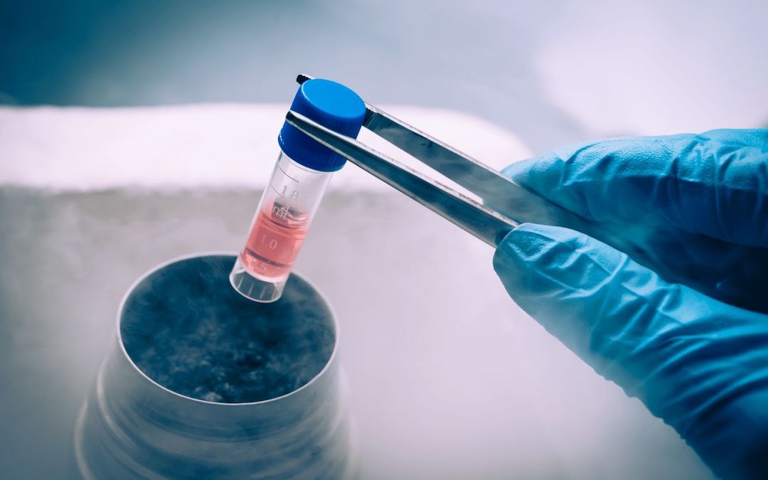
- Be cautious when freezing samples – If samples must be frozen, which can often be necessary during large cohort studies, consider using a sample type-specific freezing medium to preserve cells in their natural state and protect antibody-binding epitopes
- Check cell viability – Use a stain such as Trypan Blue to confirm sample quality before proceeding with immunostaining (when working with whole blood, it is more common for a viability stain to be included with the antibody panel)
- Resuspend cells at an appropriate density – A density of 105 – 107 cells/mL is generally recommended; using too many cells can lead to events being missed, while using too few cells can prolong run times
- Prevent cells from sticking to one another – Use a resuspension buffer that is free of Ca2+/Mg2+ and contains DNase (to digest any free DNA released from damaged cells, which can cause aggregation)
- Filter samples to remove clumps – Use an appropriately-sized cell strainer and check samples under the microscope before proceeding with the flow cytometry workflow
Supporting flow cytometry-based research
Whatever type of sample you’re analyzing for any number of markers, FluoroFinder offers a suite of solutions to simplify reagent selection and panel design. Our Antibody Search function helps you find antibodies validated for flow cytometry, while our Spectra Viewer allows you to quickly compare fluorophores from all suppliers in a single platform. Designing a panel for spectral or conventional flow cytometry? Use our Panel Builder to view 1,100+ fluorophores and 3,000,000+ antibodies from over 60 suppliers in one resource. For additional guidance, contact our support team of specialists, who are always here to help.
Sign-up for our eNewsletter to receive regular flow cytometry updates and discover the latest antibody and fluorophore offerings available for your research.


