Immunofluorescence (IF)
FluoroFinder’s tools speed up experiment design and product selection for immunofluorescence applications. Our comprehensive platform features:
- More than 3 million antibodies from all suppliers
- Optical properties of over 1,000 fluorescent dyes and proteins
- Spectra Viewer for visualization of emission spectra of multiple fluorophores excitation and emission wavelengths, and instrument laser and filter settings
Antibody Search and Immunofluorescence
Immunofluorescence is a widely used technique for the detection of cellular antigens by fluorescence or confocal microscopy. It relies on the specificity of antibody binding to the protein of interest. The fluorescence emitted by a fluorochrome, directly or indirectly conjugated to the antibody, allows visualization of the target protein under fluorescence light. The method applied to cell lines, individual cells, tissue sections, 3D-cultures, and even on entire organisms is commonly used in clinical practice and in research applications to study protein expression levels and subcellular localization. Different microscopy techniques can be used to image immunofluorescence samples.
Epifluorescence and confocal microscopy are widely used, however, super-resolution techniques, including STED (Stimulated Emission Depletion), direct stochastic optical reconstruction microscopy (dSTORM), photoactivated localization microscopy (PALM), and single-molecule FRET microscopy (smFRET) provide the highest resolution.

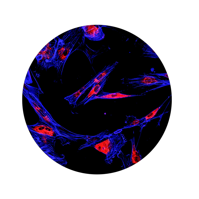
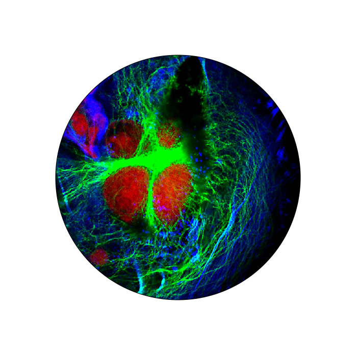
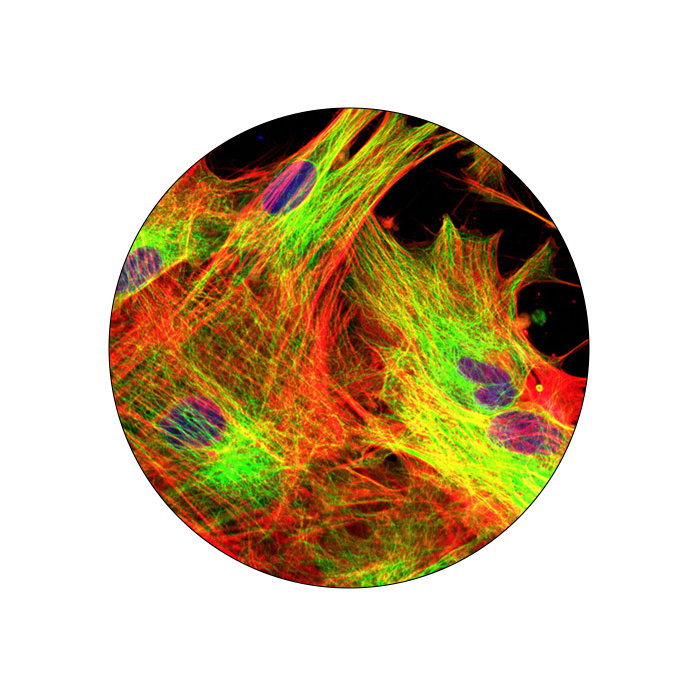
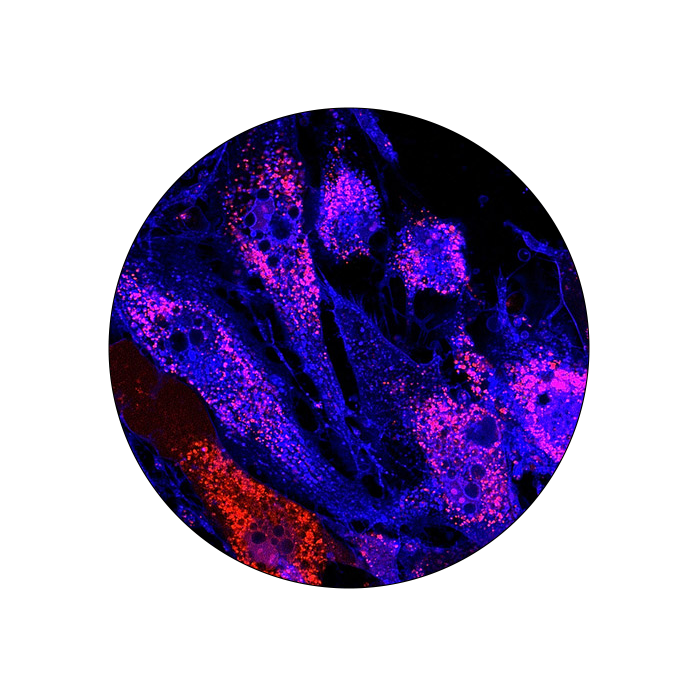
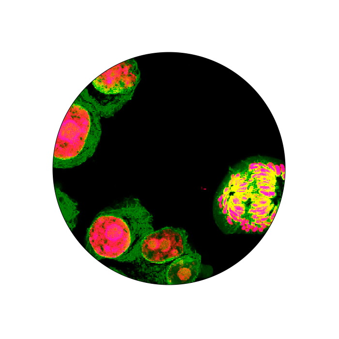
Immunofluorescence
A fixation step is essential in the preparation of samples for IF. This step uses cross-linking agents or organic solvents that immobilize target antigens without affecting the cellular architecture. It preserves the samples’ immunogenicity and prevents autolysis and protein degradation. Paraffin embedding usually follows the fixation of tissue sections. The method is used to solidify the sample for sectioning, thus allowing antibodies and dyes to easily reach the cellular compartments. Deparaffinization and antigen retrieval steps are necessary to restore antibody-antigen reactivity, especially when cross-linking reagents are used to fix the sample.
Direct Immunofluorescence
The fluorophore is directly conjugated to the primary antibody used to detect the target protein.
Indirect Immunofluorescence
A fluorescently labeled secondary antibody recognizes and binds to the primary antibody. The indirect method offers high sensitivity, signal amplification and the ability to detect multiple targets simultaneously.
References
Im K, Mareninov S, Diaz MFP, Yong WH. An Introduction to Performing Immunofluorescence Staining. ISBN 978-3540124832. OCLC 643714056.
Methods Mol Biol. 2019;1897:299-311. doi:10.1007/978-1-4939-8935-5_26 (https://www.ncbi.nlm.nih.gov/pmc/articles/PMC6918834/)


