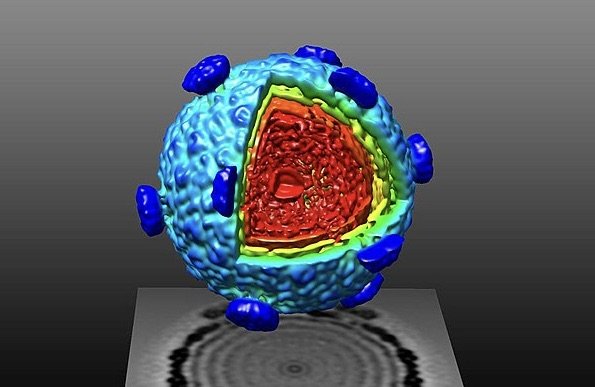
Non-conventional optics and advanced algorithms are extending the capabilities of classic microscopy.
For centuries, researchers have tried to improve the resolution and sensitivity of optical microscopy. Over time, advances in fluorescence imaging modalities have improved the level of contrast enabled researchers to overcome the Abbe diffraction limit. Recently, computational microscopy has emerged as a powerful approach to enhance the capabilities of optical microscopy. By combining principles of biomedical optics, machine learning, and algorithm design, computational microscopy offers the potential to obtain deeper insights into biological sample material.
Types of Computational Microscopy
The field of computational microscopy encompasses a steadily growing number of techniques. The following examples give a snapshot of some of the diverse strategies being employed.
Light Field Microscopy
Light field microscopy (LFM) is a 3D imaging technique first described in 2006. By inserting a microlens array into the optical path of a conventional microscope, researchers at Stanford University were able to separate light rays traveling in different directions into different camera pixels and produce a 4D light field based on intensity and angular distribution. Ray-tracing techniques were subsequently used to generate the 3D sample structure. Fourier light field microscopy (FLFM) is a recent adaptation of LFM that records the 4D light field in the Fourier domain to address common reconstruction artifacts and accelerate the reconstruction process.
Programmable Aperture Microscopy
Programmable aperture microscopy is a multi-modal technique in which a programmable light modulation device, such as a spatial light modulator or a programmable liquid crystal display, is inserted into a conventional wide-field microscope. This serves to control the distribution of light at the objective aperture plane and gives researchers the flexibility to switch between different imaging modalities (including bright field, dark field, differential phase contrast, multi-perspective imaging, and full-resolution light field imaging) without modifying any of the hardware components.
Compressed Sensing Based Microscopy
Compressed sensing is a signal processing method that can decrease the number of measurements necessary to create high-resolution images of the sample. This technique has been implemented in widefield microscopy to reconstruct images of fluorescent beads, cells, and tissues with under-sampling ratios (the ratios between the number of pixels and the number of measurements). Compressed sensing has also been adapted for conventional laser scanning microscopes by incorporating a model of point-spread function into the reconstruction framework.
Digital Holographic Microscopy
Digital holographic microscopy (DHM) is an imaging technique based on interferometry – the measurement of interference between reference and experimental waves. Instead of relying on a lens to produce an image, DHM produces a digital hologram that can be reconstructed to create an image. An advantage of DHM is that it can be used for quantitative phase imaging (QPI) – a label-free microscopy method for monitoring spatial and temporal changes in biomass.
The following are a few of the techniques developed from DHM.
- Single-Element Reflective Digital Holographic Microscopy (SER-DHM) – a cost-effective technique that uses a single one-dimensional diffraction grating for illuminating the sample and generating the digital hologram
- Speckle-Field Digital Holographic Microscopy – a method developed to reduced fixed pattern noise and improve optical sectioning capability
- Lens-Free On-Chip Holography – a compact platform with utility for point-of-care testing.
Coherent Diffractive Imaging
Coherent diffractive imaging (CDI) was first reported in 1999 as a method to image non-crystalline specimens with X-ray crystallography. Specifically, a specimen with an array of gold dots was imaged using soft (low energy) X-rays. The diffraction pattern was then oversampled and deconvoluted using an iterative algorithm. Due to CDI’s ability to improve the resolution of conventional X-ray crystallography and image larger structures, many researchers have adopted this technique for a wide range of applications. CDI has evolved into techniques including Fresnel CDI, sparsity-based single-shot subwavelength CDI, and in situ CDI, all designed to improve on the original method.
All forms of microscopy remain central to scientific discovery – from basic bright field microscopy to the most advanced computational platforms. FluoroFinder offers multiple tools for planning and executing microscopy-based research, including our Antibody Search function, Spectra Viewer, and Fluorescent Dye Directory.
Sign up for our eNewsletter to receive further updates about microscopy and many other research techniques.


