Written by Paul E. Mead, BSc (Hons), PhD, SCYM (ASCP)
Clinical Flow Cytometry: Where Measurement Meets Medicine
From Bethesda consensus to high-dimensional cytometry and the next era of clinical diagnostics
Many consequential decisions in medicine begin with a measurement. A physician makes a diagnosis, adjusts therapy, predicts prognosis, or reassures a patient based on laboratory results that must faithfully reflect biological reality. In hematological disease, clinical flow cytometry plays a central role in generating these measurements. Although the field of flow cytometry is often associated with lasers, fluorochromes, and increasingly sophisticated instruments, its true purpose goes beyond the technology itself. Clinical cytometry transforms complex cellular patterns into information that clinicians can use to guide patient care (1). Viewed through this lens, the history of clinical cytometry is more than a story of technological progress. It reflects how laboratory medicine advances when scientists, clinicians, engineers, and manufacturers align around a shared objective: translating complex biology into standardized measurements to improve patient outcomes.
For clinical flow cytometry, important moments of alignment occurred during the Bethesda Clinical Flow Cytometry Conferences, particularly the meetings held in 1995 (2-4) and 2006 (5,6). These international consensus conferences played pivotal roles in moving flow cytometry from a specialized research technique toward a standardized clinical diagnostic method. By the mid-1990s, immunophenotyping had become essential for assessing hematologic malignancies. Laboratories worldwide had developed antibody panels that identified leukemic cells with high accuracy (7,8), fundamentally changing how clinicians characterized abnormal hematopoietic cells (9). Despite these advances, considerable variability remained in how immunophenotyping was performed and interpreted (2-5). Individual laboratories designed their own antibody panels, gating strategies, and reporting conventions. While many produced excellent results, the field lacked a unified measurement framework that allowed results to be compared confidently across institutions.
The Bethesda conferences addressed this challenge directly. Rather than focusing primarily on new markers or increasingly complex panels, the discussions emphasized agreement on how immunophenotyping should be performed, interpreted, and reported so that measurements would carry consistent meaning regardless of where they were generated (2,4,5). The resulting consensus helped establish many of the foundations of modern clinical flow cytometry.
Among the most visible outcomes was the development of standardized reagent frameworks for the initial evaluation of hematologic malignancies. The 2006 conference defined consensus reagent sets for evaluating major cellular lineages, including B cells, T and NK cells, and myelomonocytic populations (5). These recommendations provided laboratories with scientifically grounded diagnostic starting points while permitting flexibility for expanded panels. Equally important were improvements in reporting practices. The Bethesda participants recommended consistent terminology for describing antigen expression and emphasized the importance of reporting fluorescence intensity distributions when clinically relevant (4,5). These changes improved communication between laboratories and clinicians and improved cross institutional comparability.
Although the technologies available at the time were less advanced than those used today, many of the concepts discussed during the Bethesda meetings, particularly the importance of standardized panels, reproducible gating approaches, and harmonized reporting, anticipated the structured, multi-institutional standardization efforts that would later emerge in initiatives such as EuroFlow (10). While many Bethesda recommendations focused on technical standardization, they also reshaped clinical practice by clarifying the indications for flow cytometric testing. Rather than focusing solely on established disease entities, the consensus emphasized symptom-driven testing and identified clinical presentations that justify the medical necessity of immunophenotyping (6). In doing so, flow cytometry became more fully integrated into routine diagnostic evaluation (11).
Another important outcome involved the professional workforce supporting the field. Clinical cytometry had grown more complex, revealing a need for specialized training. A Bethesda subcommittee recommended specialty credentials for flow practitioners. This would eventually transition the American Society for Clinical Pathology’s Qualification in Cytometry into a formal specialty certification (SCYM) (12). Certification requirements helped define professional expectations for training and expertise in the discipline.
The influence of the Bethesda consensus extended beyond laboratory procedures into the regulatory and accreditation structures that govern clinical diagnostics. Many of its recommendations were incorporated into laboratory accreditation frameworks, particularly those of the College of American Pathologists (CAP) (13). CAP flow cytometry checklists increasingly aligned with Bethesda guidance on panel design, marker selection, and validation practices. These changes strengthened quality systems within clinical laboratories. Accreditation requirements placed greater emphasis on validating antibody performance and assay sensitivity, especially for high-complexity testing. Laboratories were encouraged to perform parallel testing, share samples for rare analytes, and carefully evaluate reagent performance before adding new markers (13,14).
Rare-event detection became an increasingly important focus for clinical flow cytometry. As assays for minimal residual disease (MRD) (15-17) and paroxysmal nocturnal hemoglobinuria (PNH) (18,19) matured, laboratories had to determine analytical sensitivity and lower limits of detection (20-22). These expectations were reinforced by recommendations from the International Clinical Cytometry Society, an organization that grew from the early Bethesda collaborations.
The impact of the Bethesda meetings also extended into the regulatory environment governing clinical laboratories in the United States. Although the conferences did not directly modify federal law, they strongly influenced how flow cytometry came to be interpreted under the Clinical Laboratory Improvement Amendments (CLIA), where it is classified as high-complexity testing (14,22). CLIA regulations establish broad requirements for validation, personnel competency, and laboratory quality systems but provide relatively little discipline-specific guidance. The Bethesda recommendations helped fill that gap by defining practical expectations for training, analytical validation, and reporting practices that accrediting organizations could apply during inspections. Because accrediting organizations such as CAP adopted these recommendations into their inspection programs, the guidelines effectively became operational standards for clinical flow cytometry.
The result was a subtle but important shift. Immunophenotyping evolved from what had once been a specialized laboratory craft into a component of shared clinical diagnostic infrastructure. Once laboratories began speaking the same measurement language, new possibilities emerged. Multi‑center clinical trials could compare data with greater confidence, and risk-stratification models became more reliable. Minimal residual disease detection, where even small measurement differences can influence interpretation, became increasingly valuable in guiding treatment decisions (15-17). At the same time, survival outcomes for pediatric acute lymphoblastic leukemia improved significantly, with cure rates approaching ninety percent in many treatment protocols (23,24). Flow cytometry alone did not produce those outcomes, but it played an essential role by providing clinicians with measurements they could trust when tailoring therapy.
The importance of standardized clinical measurement in flow cytometry had, in fact, become clear earlier during the HIV/AIDS epidemic. Clinicians urgently needed reliable ways to monitor immune function in patients whose disease progression could change quickly. CD4+ T cell counts emerged as a critical biomarker for evaluating immune status and guiding treatment (25,26). Meeting this need required both innovation and practical standards, including reference materials and shared procedures to ensure consistent results across laboratories. Instrumentation improved, reagents became more consistent, and analytical methods matured. Just as importantly, the field learned a lasting lesson: clinical measurement systems require not only technological innovation but also standards, reference materials, and shared practices that ensure results remain consistent across laboratories and over time.
The years that followed the pivotal Bethesda meetings saw rapid technological expansion in flow cytometry instrumentation. Digital cytometers replaced analog systems, improving signal processing and reproducibility of fluorescence measurements. Advances in fluorochrome chemistry expanded the number of parameters that could be measured, allowing laboratories to design increasingly informative antibody panels. These developments created new opportunities for more comprehensive immunophenotypic characterization of hematologic malignancies and immune disorders.
Greater analytical capability, however, also introduced new challenges. As panel complexity increased, the need for careful panel design, standardized instrument setup, and reproducible analytical strategies became increasingly important to ensure that measurements remained clinically interpretable across laboratories. In response, collaborative international initiatives began developing more structured approaches to standardization and data interpretation. Among these was the EuroFlow Consortium, which built upon earlier consensus efforts by integrating standardized antibody panels, instrument calibration protocols, and computational analysis tools into a coordinated diagnostic framework (10,27). By combining harmonized reagents with reference databases of normal and disease-associated immunophenotypes, EuroFlow helped move the field toward a more reproducible model of cytometric diagnosis. These efforts demonstrated how coordinated collaboration among clinical laboratories, researchers, and industry partners could translate rapidly advancing technology into practical improvements in diagnostic precision.
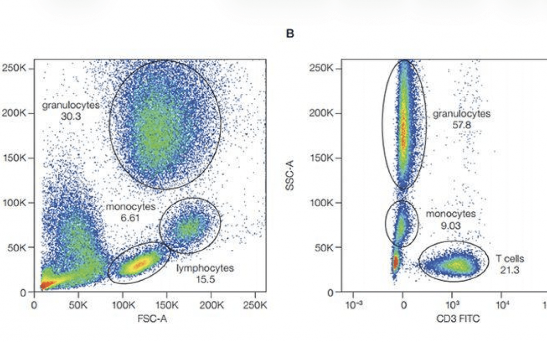
Today, clinical cytometry is again experiencing rapid technological expansion. Spectral cytometry and high-dimensional panel design have significantly increased the number of parameters that can be measured simultaneously, and these technologies are now being implemented in clinical laboratories as well as research environments (28,29). The ability to analyze dozens of markers within a single experiment provides unprecedented insight into complex immune landscapes. Rare cellular populations can be detected with remarkable sensitivity, and relationships among multiple cell subsets can be examined within a single test.
As spectral cytometry expands into clinical laboratories, an important question emerges: how can the rapidly increasing dimensionality of cytometric measurement be reconciled with the practical requirements of clinical diagnostics? Spectral platforms can now resolve dozens of parameters simultaneously, generating extraordinarily rich cellular signatures. Yet clinical diagnostics ultimately depends on measurements that are reproducible, interpretable, and comparable across laboratories. Bridging this divide will require several developments. First, greater standardization of spectral acquisition and reference controls will be necessary so that unmixing algorithms and fluorescence signatures remain stable across instruments, reagent lots, and institutions. Second, the field will need robust strategies for translating high-dimensional cytometric data into clinically interpretable metrics (30,31). While research environments can explore complex multidimensional phenotypes, clinical reporting must distill these patterns into validated diagnostic features or probability-based classifications that clinicians can apply with confidence. Finally, the analytical and computational methods supporting spectral analysis will need to mature within regulatory-grade validation frameworks comparable to those already established for conventional flow cytometry assays (14,20,21).
These considerations highlight an important reality of clinical diagnostics: the clinical laboratory environment operates under constraints that differ from those of research settings. Every assay must demonstrate not only analytical capability but also reproducibility, interpretability, and long-term stability (14). Measurements must remain consistent across reagent lots, instruments, and ideally across institutions. As panel dimensionality increases, maintaining this level of consistency becomes more challenging. Spectral cytometry relies on sophisticated unmixing algorithms and carefully maintained reference controls, and small changes in reagent performance or instrument calibration can influence high-dimensional analyses in subtle ways.
Another development shaping modern clinical cytometry is the growing use of artificial intelligence (AI) and machine learning in data analysis and laboratory quality systems. High-dimensional cytometry produces complex datasets, and computational tools can assist in identifying cell populations, recognizing patterns across large cohorts, and supporting more consistent analytical workflows (32-34). In clinical laboratories these approaches will also strengthen quality control by monitoring instrument performance, detecting shifts in fluorescence intensity, and flagging data that warrants closer review. Used appropriately, AI can complement laboratory expertise by improving analytical consistency and identifying variability earlier in the analytical process. As with any clinical technology, these tools must be validated using clear quality procedures to ensure that the measurements they support remain clinically meaningful.
These challenges are not new. Clinical cytometry has always advanced through cycles of innovation followed by standardization. New technologies expand what can be measured; the community then works collectively to ensure that those measurements remain reliable. For those working in clinical laboratories, variability rarely appears as dramatic instrument failures. More often it manifests subtly: slight shifts in antigen intensity after a reagent‑lot change, drift in instrument calibration, or small differences in gating between operators. Individually these changes may seem minor, but in assays that guide treatment decisions, even subtle changes can influence interpretation. Over time the field has learned that the greatest challenge in clinical diagnostics is not just generating data but producing results clinicians and patients can trust.
Ultimately, the purpose of clinical cytometry has always extended beyond the technologies that enable it. Instruments will continue to evolve, fluorochromes will improve, and analytical tools will become increasingly sophisticated. What remains constant is the responsibility to ensure that the measurements generated in clinical laboratories are sufficiently robust to support clinical decision making. Behind every dot plot and every gating strategy is a patient whose care may depend on the reliability of those measurements. The task for the next generation of cytometrists is therefore not simply to measure more, but to measure better and to ensure that the remarkable technologies now emerging translate into what has always defined success in clinical diagnostics: better care and better outcomes for the patients whose lives depend on our ability to get the measurements right.
About the Author
Paul E. Mead, PhD, SCYM(ASCP), is a Principal Scientist in the Department of Pathology at St. Jude Children’s Research Hospital and Technical Director of Clinical Flow Cytometry. His work focuses on clinical assay development, high-parameter spectral panel design, and translating cytometric measurement into improved diagnostic decision-making
References
- Brown M, Wittwer C. Flow cytometry: principles and clinical applications in hematology. Clin Chem. (2000) 46(8 Pt 2):1221-9.
- U.S.-Canadian Consensus recommendations on the immunophenotypic analysis of hematologic neoplasia by flow cytometry. Bethesda, Maryland, November 16-17, 1995. Cytometry. (1997) 30(5):213-74.
- Stewart CC, Behm FG, Carey JL, Cornbleet J, Duque RE, Hudnall SD, Hurtubise PE, Loken M, Tubbs RR, Wormsley S. U.S.-Canadian Consensus recommendations on the immunophenotypic analysis of hematologic neoplasia by flow cytometry: selection of antibody combinations. Cytometry. (1997) 30(5):231-5.
- Braylan RC, Atwater SK, Diamond L, Hassett JM, Johnson M, Kidd PG, Leith C, Nguyen D. U.S.-Canadian Consensus recommendations on the immunophenotypic analysis of hematologic neoplasia by flow cytometry: data reporting. Cytometry. (1997) 30(5):245-8.
- Wood BL, Arroz M, Barnett D, DiGiuseppe J, Greig B, Kussick SJ, Oldaker T, Shenkin M, Stone E, Wallace P. 2006 Bethesda International Consensus recommendations on the immunophenotypic analysis of hematolymphoid neoplasia by flow cytometry: optimal reagents and reporting for the flow cytometric diagnosis of hematopoietic neoplasia. Cytometry B Clin Cytom. (2007) 72 Suppl 1:S14-22.
- Davis BH, Holden JT, Bene MC, Borowitz MJ, Braylan RC, Cornfield D, Gorczyca W, Lee R, Maiese R, Orfao A, Wells D, Wood BL, Stetler-Stevenson M. 2006 Bethesda International Consensus recommendations on the flow cytometric immunophenotypic analysis of hematolymphoid neoplasia: medical indications. Cytometry B Clin Cytom. (2007) 72 Suppl 1:S5-13.
- Bene MC, Castoldi G, Knapp W, Ludwig WD, Matutes E, Orfao A, van’t Veer MB. Proposals for the immunological classification of acute leukemias. European Group for the Immunological Characterization of Leukemias (EGIL). Leukemia. (1995) 9(10):1783-6.
- Craig FE, Foon KA. Flow cytometric immunophenotyping for hematologic neoplasms. Blood. (2008) 111(8):3941-67.
- Maecker HT, McCoy JP, Nussenblatt R. Standardizing immunophenotyping for the Human Immunology Project. Nat Rev Immunol. (2012) 12(3):191-200.
- Kalina T, Flores-Montero J, van der Velden VH, Martin-Ayuso M, Böttcher S, Ritgen M, Almeida J, Lhermitte L, Asnafi V, Mendonça A, de Tute R, Cullen M, Sedek L, Vidriales MB, Pérez JJ, te Marvelde JG, Mejstrikova E, Hrusak O, Szczepański T, van Dongen JJ, Orfao A; EuroFlow Consortium (EU-FP6, LSHB-CT-2006-018708). EuroFlow standardization of flow cytometer instrument settings and immunophenotyping protocols. Leukemia. (2012) 26(9):1986-2010.
- Keren DF, Carey JL, Braylan RC. Flow cytometry in clinical Diagnosis. 4th ed. (2007) Chicago: ASCP Press.
- Greig B. Flow Cytometry Certification. History and future plans. International Clinical Cytometry Society e-Newsletter. (2010) Vol.1 Spring.
- College of American Pathologists. Flow Cytometry Checklist. Northfield, IL: CAP Laboratory Accreditation Program.
- Clinical and Laboratory Standards Institute. Validation of flow cytometric Immunophenotyping Assays. CLSI guideline H62. Wayne, PA: CSLI (2021).
- Coustan-Smith E, Campana D. Immunologic minimal residual disease detection in acute lymphoblastic leukemia: a comparative approach to molecular testing. Best Pract Res Clin Haematol. (2010) (3):347-58.
- Theunissen P, Mejstrikova E, Sedek L, van der Sluijs-Gelling AJ, Gaipa G, Bartels M, Sobral da Costa E, Kotrová M, Novakova M, Sonneveld E, Buracchi C, Bonaccorso P, Oliveira E, Te Marvelde JG, Szczepanski T, Lhermitte L, Hrusak O, Lecrevisse Q, Grigore GE, Froňková E, Trka J, Brüggemann M, Orfao A, van Dongen JJ, van der Velden VH; EuroFlow Consortium. Standardized flow cytometry for highly sensitive MRD measurements in B-cell acute lymphoblastic leukemia. Blood. (2017) 129(3):347-357.
- Heuser M, Freeman SD, Ossenkoppele GJ, Buccisano F, Hourigan CS, Ngai LL, Tettero JM, Bachas C, Baer C, Béné MC, Bücklein V, Czyz A, Denys B, Dillon R, Feuring-Buske M, Guzman ML, Haferlach T, Han L, Herzig JK, Jorgensen JL, Kern W, Konopleva MY, Lacombe F, Libura M, Majchrzak A, Maurillo L, Ofran Y, Philippe J, Plesa A, Preudhomme C, Ravandi F, Roumier C, Subklewe M, Thol F, van de Loosdrecht AA, van der Reijden BA, Venditti A, Wierzbowska A, Valk PJM, Wood BL, Walter RB, Thiede C, Döhner K, Roboz GJ, Cloos J. 2021 Update on MRD in acute myeloid leukemia: a consensus document from the European LeukemiaNet MRD Working Party. Blood. (2021) 138(26):2753-2767.
- Borowitz MJ, Craig FE, Digiuseppe JA, Illingworth AJ, Rosse W, Sutherland DR, Wittwer CT, Richards SJ; Clinical Cytometry Society. Guidelines for the diagnosis and monitoring of paroxysmal nocturnal hemoglobinuria and related disorders by flow cytometry. Cytometry B Clin Cytom. (2010) 78(4):211-30.
- Sutherland DR, Keeney M, Illingworth A. Practical guidelines for the high-sensitivity detection and monitoring of paroxysmal nocturnal hemoglobinuria clones by flow cytometry. Cytometry B Clin Cytom. (2012) 82(4):195-208.
- Tangri S, Vall H, Kaplan D, Hoffman B, Purvis N, Porwit A, Hunsberger B, Shankey TV; ICSH/ICCS Working Group. Validation of cell-based fluorescence assays: practice guidelines from the ICSH and ICCS – part III – analytical issues. Cytometry B Clin Cytom. (2013) 84(5):291-308.
- Wood B, Jevremovic D, Béné MC, Yan M, Jacobs P, Litwin V; ICSH/ICCS Working Group. Validation of cell-based fluorescence assays: practice guidelines from the ICSH and ICCS – part V – assay performance criteria. Cytometry B Clin Cytom. (2013) 84(5):315-23.
- Centers for Medicare & Medicaid Services. (2024). Clinical Laboratory Improvement Amendments (CLIA): Laboratory quality standards (42 CFR Part 493). Balitmore, MD: CMS.
- Pui CH, Evans WE. A 50-year journey to cure childhood acute lymphoblastic leukemia. Semin Hematol. (2013) 50(3):185-96.
- Hunger SP, Mullighan CG. Acute Lymphoblastic Leukemia in Children. N Engl J Med. (2015) 373(16):1541-52.
- Giorgi JV, Detels R. T-cell subset alterations in HIV-infected homosexual men: NIAID Multicenter AIDS cohort study. Clin Immunol Immunopathol. (1989) 52(1):10-8.
- Mandy FF, Nicholson JK, McDougal JS; CDC. Guidelines for performing single-platform absolute CD4+ T-cell determinations with CD45 gating for persons infected with human immunodeficiency virus. Centers for Disease Control and Prevention. MMWR Recomm Rep. (2003) 52(RR-2):1-13.
- van Dongen JJ, Lhermitte L, Böttcher S, Almeida J, van der Velden VH, Flores-Montero J, Rawstron A, Asnafi V, Lécrevisse Q, Lucio P, Mejstrikova E, Szczepański T, Kalina T, de Tute R, Brüggemann M, Sedek L, Cullen M, Langerak AW, Mendonça A, Macintyre E, Martin-Ayuso M, Hrusak O, Vidriales MB, Orfao A; EuroFlow Consortium (EU-FP6, LSHB-CT-2006-018708). EuroFlow antibody panels for standardized n-dimensional flow cytometric immunophenotyping of normal, reactive and malignant leukocytes. Leukemia. (2012) 26(9):1908-75.
- Nolan JP, Condello D. Spectral flow cytometry. Curr Protoc Cytom. (2013) 1:1.27.1-1.27.13.
- Ferrer-Font L, Pellefigues C, Mayer JU, Small SJ, Jaimes MC, Price KM. Panel Design and Optimization for High-Dimensional Immunophenotyping Assays Using Spectral Flow Cytometry. Curr Protoc Cytom. (2020) 92(1):e70.
- Liechti T, Weber LM, Ashhurst TM, Stanley N, Prlic M, Van Gassen S, Mair F. An updated guide for the perplexed: cytometry in the high-dimensional era. Nat Immunol. (2021) 22(10):1190-1197.
- Lee JA, Spidlen J, Boyce K, Cai J, Crosbie N, Dalphin M, Furlong J, Gasparetto M, Goldberg M, Goralczyk EM, Hyun B, Jansen K, Kollmann T, Kong M, Leif R, McWeeney S, Moloshok TD, Moore W, Nolan G, Nolan J, Nikolich-Zugich J, Parrish D, Purcell B, Qian Y, Selvaraj B, Smith C, Tchuvatkina O, Wertheimer A, Wilkinson P, Wilson C, Wood J, Zigon R; International Society for Advancement of Cytometry Data Standards Task Force; Scheuermann RH, Brinkman RR. MIFlowCyt: the minimum information about a Flow Cytometry Experiment. Cytometry A. (2008) 73(10):926-30.
- Aghaeepour N, Finak G; FlowCAP Consortium; DREAM Consortium; Hoos H, Mosmann TR, Brinkman R, Gottardo R, Scheuermann RH. Critical assessment of automated flow cytometry data analysis techniques. Nat Methods. (2013) 10(3):228-38.
- Saeys Y, Van Gassen S, Lambrecht BN. Computational flow cytometry: helping to make sense of high-dimensional immunology data. Nat Rev Immunol. (2016) 16(7):449-62.
- Ng DP, Simonson PD, Tarnok A, Lucas F, Kern W, Rolf N, Bogdanoski G, Green C, Brinkman RR, Czechowska K. Recommendations for using artificial intelligence in clinical flow cytometry. Cytometry B Clin Cytom. (2024) 106(4):228-238.


