Cell migration is fundamental to a broad range of physiological processes, including embryological development, angiogenesis, tissue regeneration, wound healing, and immune responses, to name just a few. It also has a pivotal role in the pathophysiology of conditions such as inflammatory disease and cancer. For these reasons, numerous research groups are engaged in studying migratory cell behaviors. This article looks at some of the tools and technologies available for investigating live-cell migration, with insights from Steve Titus, Senior Manager Product Management at Thermo Fisher Scientific. Methods for monitoring cell invasion and adhesion are also covered.
In Vitro Live-Cell Migration Assays and Imaging Methods
Would Healing Assay
In vitro assays for studying live-cell migration take many different forms. One of the best-known examples is the two-dimensional (2D) wound healing assay, which involves culturing cells in a microplate to form a confluent monolayer before scratching a straight line with a sterile pipette tip or stainless steel pin to simulate a wound. Various approaches are then employed to monitor the healing process.
“Wound healing assays can be visualized in real time using microscopes with environmental control capabilities, such as Thermo Fisher Scientific’s EVOS Cell Imaging Systems or the CellInsight™ CX7 LED Pro High Content Screening Platform,” explains Titus. “Alternatively, researchers can manually load, image, and unload the microplates at specified time intervals. However, a potential issue with this method is that the scratch area may shift from time point to time point depending on how the microscope stage handles and aligns the microplates.” Wound healing assays can also be based on a simple endpoint reading, whereby the cells are imaged immediately after the scratch (t=0) and incubated for a period of time (usually 3-7 days) prior to capturing a second image. % wound closure is easily calculated with the formula (a-b)/a x 100, where a is the width of the wound at t=0 and b is the width of the wound at the endpoint.
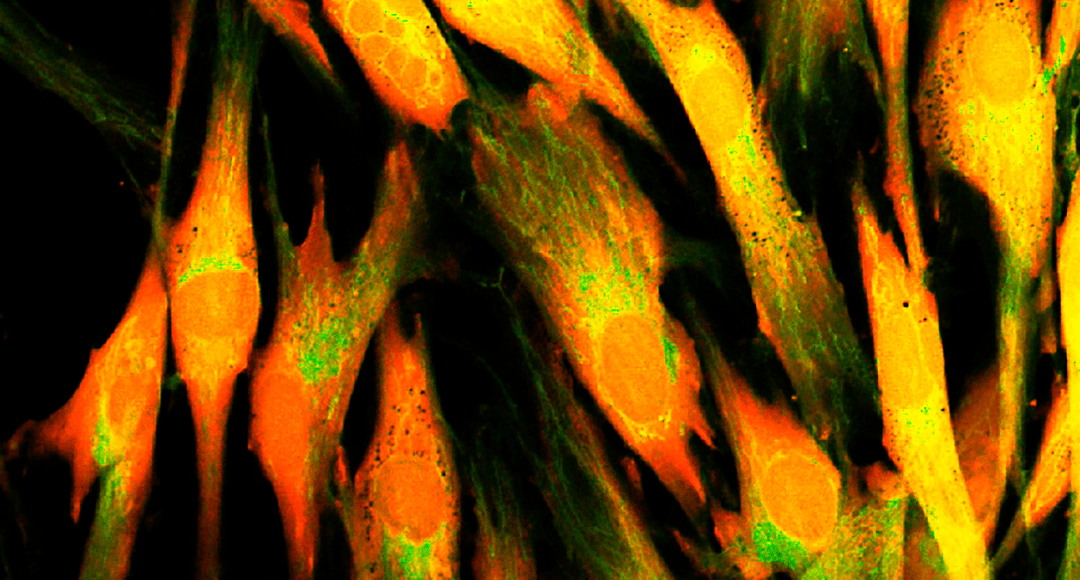
Cell Tracking Dyes
Another popular strategy for studying live-cell migration is to use cell tracking dyes for real-time monitoring of cell movement and localization. Thermo Fisher Scientific has developed two main product families for these types of studies – CellTracker™ for short-term experiments (typically 3–6 generations or 72 hours) and Qtracker™ for long-term tracking (typically 6–10 generations or 5–14 days). “Our Cell Tracking Dyes are available in a range of fluorescent colors to maximize experimental flexibility,” says Titus. “They work by entering live cells, where they are retained through multiple cycles of cell division without being transferred to neighboring cells.”
Transwell Inserts
Live-cell migration assays may also be performed using transwell inserts, which come with different pore sizes and surface treatments. A typical experiment involves applying cells to the inserts, which are suspended in microplate wells that have been pre-loaded with a buffer containing chemoattractant. Following incubation, non-migrated cells are removed from the upper surface of the transwell insert membrane with a cotton-tipped applicator, then migrated cells on the lower surface are fixed and stained1. Staining can be either colorimetric, using dyes such as Crystal Violet or Hematoxylin, or fluorescent, using DAPI or labeled antibody reagents.
Live-Cell Invasion Assays for Studying Cancer Metastasis
Live-cell invasion assays are widely used for studying cancer metastasis, which is where cells originating from a primary tumor spread through the circulatory and lymphatic systems before crossing basement membranes and endothelial walls to colonize distant organs. Early methods for studying live-cell invasion involved the laborious process of isolating basement membranes from structures such as the amnion, but the development of Matrigel® (a solubilized basement membrane extract) in the mid-1980s represented an important breakthrough for performing these types of experiments2,3.
In a seminal 1987 publication, Albini et al. reported the use of Matrigel-coated filters in the well-established Boyden chamber assay4,5. Critically, while malignant cell lines could cross the matrix, non-tumorigenic cell lines such as human fibroblasts and mouse 3T3 cells could not. Additionally, when the invaded cells were injected into nude mice, in vitro invasion was found to correspond to relative metastatic potential in vivo. This type of invasion assay is still in use today, although early Boyden Chamber devices have been superseded by modern microplates and Matrigel-coated transwell inserts.
Cell Adhesion Assays and Adhesion Molecules
Cell adhesion is involved in stimulating signals that regulate the cell cycle, cell differentiation, cell migration, and cell survival. As such, it facilitates a vast array of physiological processes. Changes in cell adhesion are implicated in diseases like arthritis, osteoporosis, and atherosclerosis, and loss of cell-to-cell adhesion is one of the hallmarks of cancer.
Cell adhesion molecules (CAMs) are a major focus of research aimed at revealing the underlying mechanisms of cell adhesion. They are classified into four main families, all of which have different functions. Cadherins localize to adherens junctions and interact with like cadherins on adjacent cells to form barriers and mediate cell polarization. Integrins link the actin cytoskeleton of a cell to external structures and are important for signal transduction. Selectins facilitate transient cell-cell interactions, most notably in the bloodstream, with leucocyte rolling on vascular surfaces being a prime example. The immunoglobulin superfamily encompasses a diverse set of cell-cell contact proteins, including vascular and neural cell adhesion molecules (VCAM and NCAM), intercellular adhesion molecules (ICAMs), and Nectins.
Methods for studying cell adhesion include static/passive assays, in which cells are allowed to settle onto a substrate, and flow-based/dynamic assays, which involve cell perfusion. A well-cited example of the former describes the use of a surface coated with human mesenchymal stem cells (hMSC) to demonstrate the importance of CD44 in the interaction between hMSC and hematopoietic progenitor cells, which was significantly reduced by administration of a monoclonal CD44 antibody6. A recently reported technique, termed motion blur microscopy, provides an easy to implement means of imaging cell interactions in microfluidic channels during whole blood flow7. Besides these methods, countless bespoke assays have been developed by researchers to establish roles for distinct proteins in cell adhesion.
Supporting Your Research
Selecting compatible fluorescent dyes can be challenging, particularly in multiplexed experiments. If you’re using fluorescent reagents such as cell tracking dyes or fluorophore-labeled antibodies for studying cell migration, invasion or adhesion, FluoroFinder offers a suite of tools that can simplify experimental design. Leverage our Fluorescent Dye Directory for detailed information on the optical properties and spectral profiles of more than a thousand fluorochromes, and use our Spectra Viewer to identify the right dyes for your instrument’s laser and filter configurations.
You can also find a wealth of resources on Thermo Fisher Scientific’s website, including a comprehensive Guide to Live-Cell Imaging Reagents that encompasses the Stain-iT Cell staining Simulator tool to select dyes based on live/fixed status.
References:
- Justus CR, Marie MA, Sanderlin EJ, Yang LV. Transwell In Vitro Cell Migration and Invasion Assays. Methods Mol Biol. 2023;2644:349-359.
- Albini A. Tumor and endothelial cell invasion of basement membranes. The matrigel chemoinvasion assay as a tool for dissecting molecular mechanisms. Pathol Oncol Res. 1998;4(3):230-241.
- Passaniti A, Kleinman HK, Martin GR. Matrigel: history/background, uses, and future applications. J Cell Commun Signal. 2022;16(4):621-626.
- Chen, HC. (2005). Boyden Chamber Assay. In: Guan, JL. (eds) Cell Migration. Methods in Molecular Biology™, vol 294. Humana Press.
- Albini A, Iwamoto Y, Kleinman HK, et al. A rapid in vitro assay for quantitating the invasive potential of tumor cells. Cancer Res. 1987;47(12):3239-3245.
- Wagner W, Wein F, Roderburg C, et al. Adhesion of human hematopoietic progenitor cells to mesenchymal stromal cells involves CD44. Cells Tissues Organs. 2008;188(1-2):160-169.
- Goreke, U., Gonzales, A., Shipley, B. et al. Motion blur microscopy: in vitro imaging of cell adhesion dynamics in whole blood flow. Nat Commun 15, 7058 (2024).


