Written By Sarah Locknar
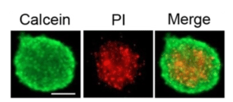
Cover: Spheroids labeled with Calcein-AM (green) for live cells and propidium iodide (PI) for dead cells imaged with widefield fluorescence microscopy. Scale bar = 100 micron Used under CC by 4 license without modification from [1] Figure 3A.
Organoid and spheroid analysis
Basic science and pharmaceutical research have been moving away from animal testing for many years. Traditional 2-D cell culture (on bare or coated slides orbut dishes) is useful for early-stage screening experiments, but for studies of toxicity and efficacy in later stages of drug development, 2-D cultures are too different from whole animals to provide meaningful data. Regulatory agencies are actively encouraging the development of non-animal models (NAMs) to replace slow, costly and ethically questionable animal studies, especially for monoclonal antibody drugs, where the human and animal responses can be very different. NAM 3-D cultures are not only more specific to humans (often grown from primary human cells), their use enables fast, high-throughput, semi-automated analysis.
The simplest 3-D cell cultures are called spheroids. These particles are usually composed of a single cell type (or a small number) that spontaneously form clusters. They are quite uniform, spherical and fragile, relying solely on cell-to-cell adhesion for structural integrity. Because of their size, they can settle out of solution without gentle rocking or agitation. Larger spheroids can become hypoxic in the center, causing necrosis (cover image).
The next most complicated 3-D cell culture is the organoid. Organoids are grown from stem cells in a gel (Matrigel, collagen, etc.) attached to the surface of the plate (Figure 1). Growth factors and other signaling molecules in the culture medium induce cells to differentiate into many of the cell types present in the original tissue. The gel more closely mimics an in vivo environment and lends some structural integrity to isolated organoids.
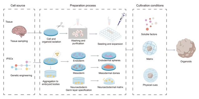
Figure 1. Organoid construction process. Organoids can be generated from tissue-derived cells or induced pluripotent stem cells. Tissue samples are obtained, washed, and cut into small fragments, followed by enzymatic digestion to isolate individual stem cells. After washing and purification, the harvested stem cells are used to seed and generate organoid cultures. Used under CC by 4 license without modification from [2] Figure 1.
Unlike spheroids, organoids exhibit substantial heterogeneity in size, shape and cell type, even when grown in identical conditions (Figure 2). Some are solid, some have lumens, budding, folds, etc. Size often correlates with functional properties, so size sorting can be important for longitudinal analysis. [3] Similar to spheroids, organoids can become hypoxic in the center when they get too large. Tissue and tumor organoids are also often co-cultured with immune organoids to more closely simulate immune responses in-vivo. [3] For comprehensive reviews of current organoid engineering, see references [4] [2]. The Human Cancer Models Initiative (HCMI) maintains primary patient-derived organoid models with additional data (clinical diagnosis, patient age, etc.) that can be ordered from ATTC.
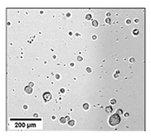
Figure 2. Size and shape variation of organoids. Used under CC by 4 license without modification from [3] Figure 3a.
The most complicated 3-D cell culture is the organ-on-a-chip (OOC). These multi-cell tissue cultures are grown in a microfluidic chamber that can mimic nutrient flow and pressures present in intact tissues. Organoids have also been grown inside of microfluidic chambers to gain similar benefits during their growth and development.
Monitoring and Analysis of Spheroids and Organoids
Approaches to monitoring the health of 3-D cultures include indirect monitoring of media, dissociation followed by flow cytometry, gentle sorting of live or fixed 3-D particles, and longitudinal imaging studies.
Indirect methods for 3-D culture monitoring
The culture media can be monitored for changes in pH, metabolic and/or other biomarkers. For spheroids, 3-D particles are allowed to settle (or are gently centrifuged) before aspirating the media, whereas media over organoids grown in a matrix is simply aspirated. Matrix-only controls should also be collected to account for effects of their complex composition. OOCs with a fluid channel, can be monitored continually if desired. Media samples can be used in multiplexed ELISA-based assays to detect soluble proteins and peptides, including cytokines, angiogenesis factors and matrix metalloproteinases. Colorimetric or luminescence assays can be used to detect glucose and lactic acid levels over time, while mass spectroscopy can characterize low-molecular-weight metabolites and essential amino acids.
It is well known that 3D cell cultures release a large number of extracellular vesicles (EVs) which can provide a rich source of additional information. EVs can be concentrated and characterized by electron microscopy or light scattering to determine size, labeled with fluorescent markers and characterized by flow cytometry and their contents can be analyzed using RNA sequencing or mass spectroscopy for lipidomic and proteomic information.
Destructive analysis of spheroids and organoids
3-D cultures can be dissociated into individual cells and characterized using flow cytometry or lysed and characterized biochemically, but valuable structural information is lost using these methods. Another option is to fix and stain the 3-D cultures for analysis by microscopy or flow cytometry. Most modern protocols today try to preserve 3-D structure.
Gentle sorting of intact spheroids and organoids
The aggregation and growth methods used in 3D cultures often lead to undesirable variations in size, shape and composition between particles. Selecting a similar subpopulation of particles can be beneficial before proceeding with other studies. Spheroids tend to be more fragile than organoids and should be harvested with care. Capillary flow or automated systems [5] preserve the structure better than high-shear manual pipetting. An automated Pick-Flow-Drop technique can be used instead of flow cytometry to select particles of a particular size for isolation and longitudinal analysis. [5] Droplet microfluidics have been employed to encapsulate spheroids in hydrogel matrices to increase structural integrity for sorting and later growth. [6]
Sorting 3D particles in a flow cytometer requires special treatment. If sorted particles are to be used for future longitudinal studies (in microplates for instance), light scattering, intrinsic fluorescence (of metabolites like NAD(P)H and FAD), or non-toxic vital dyes should be used to maximize cell viability. Typical flow cytometers use high pressure (with high shear forces) to increase throughput, which can easily break apart 3-D particles. Commercially available 3-D particle sorters use large nozzles and low pressure to balance throughput with gentleness. The Union Biometrica COPAS adds a high-viscosity sheath liquid to stabilize laminar flow and reduce shear, while Cellenion spheroONE uses acoustic waves to isolate and dispense particles without a sheath liquid. Imaging during sorting is important, not only for size gating, but to characterize other features of organoids (presence or absence of a lumen, cellular composition, overall shape, etc.) and to remove debris. The COPAS offers brightfield imaging with total fluorescence signal detection (up to 4 lasers and 12 detectors) and the spheroONE offers both brightfield and fluorescence imaging.
Longitudinal imaging of spheroids and organoids
Once the 3D particles are dispensed into microplates, high-resolution longitudinal imaging can be used to see how they react to drugs or other agents. Smaller particles (< 60 micron) can be monitored using phase or brightfield, widefield fluorescence and spinning disk (confocal) microscopy, using standard microscopes or multimode microplate readers. Microplate readers have the advantage of high-throughput capabilities, with automated plate handling, precision positioning motors, environmental control, and the ability to image at low and high magnification. In fact, precision motors and plate indexing software can enable longitudinal particle studies without a separate particle sorting step. The Agilent BioTek Cytation C10 has LED-based wide-field fluorescence, and spinning disk confocal (6 laser options) with brightfield and phase contrast capabilities. Molecular Devices offers the ImageExpress HCS.ai with transmitted light, widefield fluorescence and LED (5-color) or laser (7-color) based spinning disk confocal, designed to work with their CellXpress.ai Automated Cell Culture System.
Disadvantages of spinning disk systems include phototoxicity during longitudinal studies and the limited imaging depth of these systems, which requires particles to be positioned close to the bottom of the well. Multiplate readers based on multiphoton confocal sectioning are not currently available, but low throughput manual imaging with a multiphoton microscope is always an option for large particles. Optical coherence tomography has also been used to image larger organoids. [7] Another option is light-sheet microscopy as implemented in Hamamatsu’s CYTOQUBE® microplate imager using Zyncscan® technology. A vertical sheet illuminates the sample well, with oblique fluorescence imaging of the (~ 6 x 0.4mm) XZ plane. The stage moves the microplate over the light sheet to give vertical slices through the sample (fluorescence tomography) with up to 4 excitation lasers. Resolution is 2.75 x 2.75 x 6.215 microns (X,Y,Z). It does not take brightfield images and can only process a single plate at a time, but this technology is one to watch.
Other consideration
Working with 3D particles shares many of the same problems as tissue analysis. Dye penetration and imaging through thick, dense samples are always tricky and longitudinal imaging studies generate large, 3-D, multicolor datasets that need to be processed, stored and analyzed. AI data processing is becoming popular for automated cell counting, alive/dead analysis and growth kinetics. [3] [4] Standardized protocols for image analysis are still being developed, but imaging, combined with indirect monitoring, leads to very detailed descriptions of the effects of drugs or other agents on the 3-D particles. Overall, the use of spheroids and organoids is reducing the burden on animal testing in basic science and drug development, and accelerating the translation of medical research into clinical applications for the benefit of us all.
References
| [1] | Lee, 2018. doi:10.7150/thno.26439. |
| [2] | Cheng, 2026. doi:10.1186/s43556-026-00422-7. |
| [3] | Ferreira, 2025. doi:10.1007/s13402-024-00958-2. |
| [4] | Kim, 2026. doi:10.1038/s44385-025-00054-6. |
| [5] | Zieger, 2024. doi:10.1002/adhm.202303350. |
| [6] | Ezzo, 2025. doi:10.1021/acsbiomaterials.5c01015. |
| [7] | Fei, 2022. doi:10.3390/bioengineering9030121. |